- Jul 8
How to Choose the Right Needle for Radiofrequency Thermocoagulation: A Clinician’s Guide
- Haroun Gajraj
- Thermocoagulation
By Dr. Haroun Gajraj | VeinCare Academy | 8th July 2026
Written by Dr. Haroun Gajraj, GMC-registered Vascular Specialist, Founder & Board Member of the British Association of Sclerotherapists (BAS).
This article is written for clinicians who perform, or are learning to perform, radiofrequency thermocoagulation (shortwave diathermy at 4 MHz) for facial and leg telangiectasia. It covers how electrolysis needles are classified, what the letters and numbers on the packaging actually mean, the clinical rationale for insulated versus non-insulated designs, and the evidence—such as it is—behind current practice recommendations.
🎥 Prefer video? Watch the full review on YouTube: "How to Choose the Right Needle for Radiofrequency Thermocoagulation (F3 vs K3 vs Insulated)" [→click here]
📋 Quick Summary for Clinicians
Shank Size (F vs. K): Dictates machine compatibility only; has zero clinical impact on treatment outcome. Match to your specific handpiece.
Shaft Size (F2–F6): The critical clinical variable. Match the probe diameter to the vessel diameter. F3 (0.003") is the standard "Goldilocks" size for facial telangiectasia.
Insulation Status: Insulated needles concentrate thermal energy strictly at the exposed tip, protecting the epidermis, reducing tissue trauma along the dermal track, and minimizing the risk of post-inflammatory hyperpigmentation (PIH) in darker skin tones.
Material Claims: No indexed comparative clinical trials support the superiority of gold-coated needles over standard stainless steel. Focus procurement choices on size and insulation.
Contents
Introduction
Probe anatomy: the four sections of an electrolysis needle
What is the difference between F and K shank needles?
The shaft number: the variable that actually matters clinically
Clinical performance: insulated vs. non-insulated electrolysis needles for vascular work
Gold needles: what the evidence does and does not show
Facial versus leg telangiectasia: matching probe to vessel and modality
A practical needle selection framework
Key clinical points
Frequently Asked Questions (FAQs)
References
1. Introduction
If you perform radiofrequency thermocoagulation of telangiectasia, you will have faced this moment: staring at a needle catalogue trying to work out whether to order the F3, the K3, the insulated version, or the gold one—and wondering whether the choice actually matters.
It does matter, and the reasoning is worth understanding properly. But the volume of contradictory advice in circulation, much of it borrowed from the electrolysis-for-hair-removal world rather than from modern vascular practice, makes this harder than it should be.
This article sets out to clarify the classification system, explain the electrosurgical physics, and give a practical rationale for needle selection in telangiectasia work—while being honest about where the clinical evidence runs out.
One important caveat applies from the outset. Unlike Microsclerotherapy, where several randomised trials and formal international guidelines address treatment decisions in detail, probe selection for radiofrequency thermocoagulation of telangiectasia is not covered by any current guideline recommendation from the ESVS [5], SVS/AVF/AVLS, or the International Union of Phlebology. The Cochrane systematic review on treatment of telangiectasia and reticular veins does not address the question [1]. What follows is therefore a synthesis of published clinical and histological evidence, electrosurgical physics, and accumulated practice—not a summary of high-grade comparative trial data. That distinction matters and will be stated explicitly throughout.
2. Probe Anatomy: The Four Sections of an Electrolysis Needle
Before discussing classification systems, it is useful to have a clear mental model of the probe itself.
Every standard electrolysis needle has three or four distinct sections depending on its construction.
One-piece needles have four sections: the shank, the taper, the shaft, and the tip. Two-piece needles—such as the Sterex range used with the Sterex and Silhouette machines—omit the taper; the hub connects directly to the shaft, giving three functional sections.
The shank is the thick proximal end that plugs into the handpiece. Its sole function is mechanical: it determines whether the needle fits your machine. It has no effect on treatment outcome whatsoever.
The taper (one-piece designs only) is the transitional zone between the shank and the shaft. It provides structural continuity but has no independent clinical role.
The shaft is the long, thin functional section that carries current to the treatment site. The diameter of the shaft is the primary determinant of how the needle performs clinically. It governs current density, heat concentration, and the size of vessel the probe is best suited to. This is the part of the classification that matters for clinical decision-making.
The tip is the micro-polished distal point that contacts or enters the skin surface and the vessel wall. In insulated needles, the tip is the only exposed conductive segment; the shaft above it carries an insulating coating up to, but not including, the tip itself. In non-insulated needles, the full shaft is conductive.
Understanding this anatomy makes the rest of the classification system immediately legible.
3. What is the Difference Between F and K Shank Needles?
The letter prefix in an electrolysis needle designation—F or K—describes the diameter of the shank.
An F-shank is 1.25 mm in diameter. A K-shank is 0.8 mm. That is the only difference between them.
An F3 and a K3 needle, used in the same patient at the same current setting, produce identical treatment results. The shaft diameter is the same, the heat distribution is the same, and the clinical outcome is the same. The letter tells you which machine the needle fits, and nothing else.
In the UK, the majority of machines—including the Sterex and Silhouette platforms—use F-shank needles. K-shank is more common in North American machines, including VeinGogh. If someone asks you whether they should use F or K, the answer is whichever fits your handpiece. There is no clinical argument for either.
4. The Shaft Number: The Variable That Actually Matters Clinically
The number following the letter designates the shaft diameter, measured in thousandths of an inch.
The core principle is straightforward: match the shaft diameter to the vessel size. A thinner shaft concentrates more energy at the tip—the same principle as forcing water through a narrower tube at the same pressure. A needle that is too fine for the target vessel will run excessively hot and is harder to control; one that is too large for the target distributes heat across a broader zone than necessary.
The clinically relevant size range for telangiectasia work is:
F2 (0.002 inches / 0.05 mm): The finest standard needle. It runs hot at a given current setting and requires experience and conservative current settings to avoid overtreatment. Technically useful for the finest facial vessels in experienced hands.
F3 (0.003 inches / 0.075 mm): The clinical standard for facial spider veins. This is the shaft size used in the majority of dedicated RF thermocoagulation platforms and recommended by virtually every professional training course for facial telangiectasia. It balances precision with practical handling characteristics—the clinical "Goldilocks" for facial vein work.
F4 (0.004 inches / 0.1 mm): Slightly larger with a somewhat broader heat spread. Useful for larger facial vessels, for leg work where vessels are somewhat bigger, or for patients who are particularly pain-sensitive.
F5 and F6 (0.005–0.006 inches / 0.125–0.15 mm): More suited to leg telangiectasia in the 0.2–0.5 mm diameter range, where the larger vessel size calls for proportionately more energy delivery.
Histological work on ohmic thermolysis supports the broad principle of size matching, reporting greater than 90% clearance in telangiectasia under 0.5 mm in diameter [4]. This is not a randomised comparison of shaft sizes, but it does support the mechanistic principle that fine probes achieve effective vessel destruction in fine vessels when used carefully.
5. Clinical Performance: Insulated vs. Non-Insulated Electrolysis Needles for Vascular Work
This is the part of needle selection that generates the most debate, and where the conventional advice most warrants scrutiny.
The conventional position
The traditional advice—still repeated in some training materials—is that non-insulated needles should be used for spider vein work, and that insulated needles are for hair removal. The reasoning given is that for surface thermocoagulation, heat along the full shaft is acceptable because the target vessel is at or near the skin surface, making precise depth targeting unnecessary.
Why that reasoning deserves challenge
When treating a facial telangiectasia, the target is the vessel wall. That vessel sits below the skin surface, typically 0.1–0.5 mm into the dermis. The needle tip must penetrate the epidermis to reach it. Thermal energy needs to be concentrated at that tip, at the vessel wall—not distributed along the shaft in contact with the overlying skin.
The diagram below, taken from the companion video slides, illustrates the difference directly.
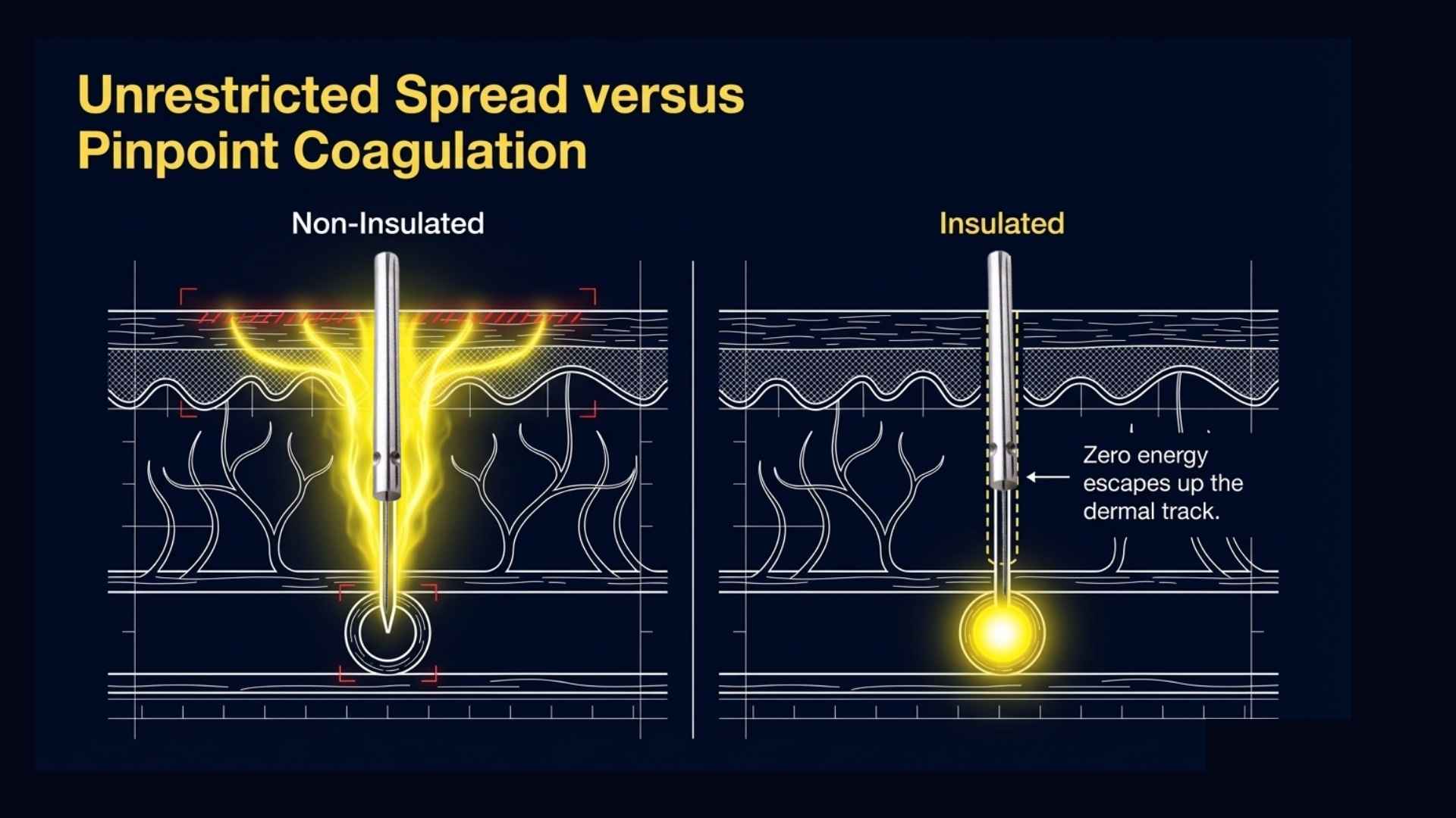
Left panel (Non-Insulated): heat radiates broadly along the full inserted shaft, spreading into the overlying epidermis and surrounding dermis as well as the target vessel. Right panel (Insulated): zero energy escapes up the dermal track; all thermal energy is concentrated at the exposed tip, precisely at the vessel wall.
A non-insulated needle produces heat along its full conductive length. Some of that energy is therefore deposited into the overlying epidermis and superficial dermis—the tissue the clinician is trying to protect. With repeated passes, the clinical consequence is an increased risk of surface heating, crusting, and pigment change.
An insulated needle—where the shaft carries an electrical insulating coating right up to, but not including, the exposed tip—concentrates all thermal energy at that distal point. The result is more precise delivery to the vessel wall and materially less collateral heat spread to surrounding tissue.
The Physics of Insulated vs. Non-Insulated Needles
From a pure electrosurgical physics standpoint, the insulation acts as a dielectric barrier. By restricting the conductive surface area to the minute exposed tip, the current density (J = I/A) is drastically increased for any given current (I). This allows the clinician to achieve the required thermal closure temperature at the vessel wall using less total energy output, significantly minimizing collateral thermal damage.
Insulated Needles in Commercial Platforms and Darker Skin Tones
This is not a fringe position. The insulation argument is the engineering logic built into the major dedicated commercial RF thermocoagulation platforms. Thermavein, Veinwave, and VeinGogh all use insulated needle designs as a core component of their systems. Sterex themselves note that insulated needles are particularly valuable for patients with Fitzpatrick skin types 3–6, where confining current to the tip reduces the risk of collateral epidermal thermal damage and the associated risk of post-inflammatory hyperpigmentation (PIH). That is a clinically important consideration for any practice treating patients with darker skin tones.
Comparing the two designs: clinical dynamics
Clinical Dimension Non-Insulated Needles Insulated Needles (e.g., Sterex F3i) Energy Conduction Broad; travels along the entire inserted metal shaft Pinpoint; strictly limited to the exposed tip Tissue Trauma Risk Higher potential for collateral epidermal heating Minimised surface trauma; protects the dermal track Handling Profile Extremely smooth piercing due to specialised tapered point Controlled, familiar feel ideal for delicate facial networks
This framing clarifies that the choice between insulated and non-insulated is not simply about efficacy at the vessel. It is also about where else energy goes, and what the cost of that spread is to the surrounding tissue.
Where the conventional advice appears to come from
The recommendation to use non-insulated needles for vascular work appears to have been imported wholesale from electrolysis-for-hair-removal practice, where the clinical priorities are genuinely different. In hair removal, the target follicle runs deep and the needle tracks it along a natural channel; distributing heat along the shaft may be useful for that application. In telangiectasia treatment, the vessel is superficial and the clinical priority is protecting the overlying epidermis while delivering targeted energy to the vessel wall. The two applications are not equivalent, and the transferred advice may not be appropriate.
What the clinical evidence actually says
The direct published evidence for this debate is weak. An older technical paper on insulated electrosurgical needles reported satisfactory results using a partially insulated design, but it was not a modern controlled comparison with standardised endpoints [3]. There is no published randomised controlled trial, and no adequately powered head-to-head prospective study, comparing insulated with non-insulated needles for thermocoagulation of facial or leg telangiectasia.
The recommendation for insulated needles therefore rests on electrosurgical physics, engineering consensus, and accumulated clinical experience—not on Level 1 or 2 evidence. That caveat applies with equal force to the recommendation for non-insulated needles. Neither position is supported by a comparative trial. The difference is that the insulation argument has the more coherent mechanistic rationale and is consistent with how the leading commercial thermocoagulation platforms are designed and marketed.
6. Gold Needles: What the Evidence Does and Does Not Show
Gold-coated needles are widely marketed for telangiectasia treatment, sometimes with the suggestion that gold is superior to standard stainless steel in terms of efficacy, patient comfort, or cosmetic outcome.
The published clinical evidence does not support this claim. No indexed comparative study demonstrates that gold needles improve efficacy, safety, pain experience, or cosmetic outcome in radiofrequency thermocoagulation of facial or leg telangiectasia. The variables that have a demonstrable mechanistic relationship to treatment outcome—shaft diameter, active exposed tip length, insulation design, and calibre matching to the target vessel—are all independent of whether the metal is gold or stainless steel.
This is an important negative finding. In the absence of trial evidence, premium material claims for gold needles should be treated with appropriate scepticism. The more important procurement questions are shaft size, insulation status, and shank compatibility with your machine.
7. Facial Versus Leg Telangiectasia: Matching Probe to Vessel and Modality
The needle selection principles described above apply primarily to facial telangiectasia and to the finest leg spider veins—those typically at or below approximately 0.3 mm in diameter.
For leg telangiectasia, the context is more complex. Vascular electrosurgery and thermocoagulation are best reserved for the finest superficial vessels: those where surface sclerotherapy is least reliable, typically below approximately 0.3 mm where the vessel is too fine to accept sclerosant intraluminally with confidence. For these vessels, an F5 or F6 shaft is generally more appropriate than the F3 used for facial work, because the target vessel is somewhat larger and requires proportionately more energy delivery.
Where thermocoagulation is used alongside Microsclerotherapy in the same session for leg telangiectasia, a randomised pilot study found that adjuvant RF thermocoagulation improved cosmetic outcomes and reduced hyperpigmentation compared with sclerotherapy alone [2]. The two modalities address different parts of the size spectrum and are complementary rather than competing.
Larger leg vessels—blue telangiectasia above approximately 1 mm, reticular feeders, or vessels with demonstrable haemodynamic reflux—are generally better treated by Microsclerotherapy or by addressing the underlying venous network first. Escalating probe size to extend thermocoagulation into lesions that belong to a different modality is not appropriate clinical practice. The modality decision should precede and drive the probe decision, not the other way around.
8. A Practical Needle Selection Framework
Based on the evidence and reasoning described above, the following framework reflects current practice for clinicians performing RF thermocoagulation with an F-shank machine.
-
For facial telangiectasia:
F3 insulated (e.g., Sterex F3i) is the practical default for most facial telangiectasia work. It balances precision, manageable heat characteristics, and epidermal protection.
F2 insulated is an option for the finest facial vessels in experienced hands, but requires conservative current settings and careful technique.
F4 insulated may be preferred for larger facial vessels or pain-sensitive patients.
If skin penetration is technically difficult, a specialised ultra-sharp probe such as the Ballet TEL is an appropriate alternative. Its penetration characteristics address a different technical problem from insulation, and the two are not mutually exclusive considerations.
-
For leg telangiectasia (thermocoagulation-appropriate size range):
F5 or F6 is generally more suited to vessels in the 0.2–0.5 mm range where thermocoagulation is indicated.
Match shaft size to vessel size as the primary decision.
-
For Fitzpatrick skin types 3–6:
Insulated needle designs have a particularly strong rationale for reducing the risk of collateral epidermal heat and post-inflammatory hyperpigmentation.
-
Shank selection:
Use whichever shank fits your machine. F-shank for Sterex and Silhouette platforms in the UK; K-shank for VeinGogh and certain North American systems.
-
Gold versus standard:
No clinical basis exists for preferring gold-coated needles. Shaft size and insulation status are the variables that matter.
9. Key Clinical Points
The letter prefix (F or K) indicates shank diameter and machine compatibility only. It has no effect on treatment outcome.
The shaft number is the clinically important variable. It determines current density, heat concentration, and the size of vessel the probe is best suited to.
F3 (0.075 mm shaft) is the practical standard for facial telangiectasia and is used by the majority of dedicated commercial RF thermocoagulation platforms.
Insulated needles concentrate thermal energy at the exposed tip rather than along the full shaft, providing a mechanistically coherent rationale for reduced epidermal heat spread and minimised tissue trauma along the dermal track.
The major commercial thermocoagulation platforms—Thermavein, Veinwave, and VeinGogh—all use insulated needle designs. The conventional advice to use non-insulated needles for vascular work appears to derive from hair removal practice, where the clinical priorities differ.
No randomised trial compares insulated with non-insulated needles for telangiectasia thermocoagulation. The insulation recommendation rests on electrosurgical logic, engineering consensus, and clinical experience—not on Level 1 evidence. The same caveat applies symmetrically to the non-insulated recommendation.
No clinical evidence supports the use of gold-coated needles over standard stainless steel for thermocoagulation of telangiectasia. Shaft size and insulation status are the variables that matter.
For leg telangiectasia, thermocoagulation is best reserved for the finest vessels where Microsclerotherapy is least reliable. Larger leg vessels and feeders remain the domain of Microsclerotherapy, not of escalated probe size.
10. Frequently Asked Questions (FAQs)
What happens if I use an F-shank needle in a K-shank machine?
It simply will not fit. The F-shank has a wider diameter (1.25 mm) than the K-shank holder (0.8 mm). Forcing it will damage the handpiece collet. Always verify your machine's handpiece requirements before ordering.
Can using an insulated needle decrease patient pain during treatment?
While pain is subjective and not formally compared in clinical trials for this application, limiting the thermal energy to the targeted vessel wall reduces unnecessary surface burns along the dermal track. Many clinicians report that patients find insulated needles more tolerable due to reduced epidermal collateral heating.
Why do some training manuals still insist on non-insulated needles for vascular work?
Most early surface electrosurgical protocols were adapted directly from cosmetic electrolysis manuals written for permanent hair removal. In hair removal, heating the entire hair follicle shaft can be advantageous. However, for vascular telangiectasia, preserving the overlying skin while sealing the vessel requires precise depth targeting, making insulation mechanistically superior.
Does shaft size alter the electrical power output setting on my RF device?
Yes. A finer needle (like an F2) concentrates the electrical current onto a much smaller surface area, drastically increasing current density. You will generally need to lower your machine’s power or duration settings when switching from an F4 or F3 to an F2 to prevent overheating the tissue.
11. References
Nakano LCU, Cacione DG, Baptista-Silva JCC, Flumignan RLG. Treatment for telangiectasias and reticular veins. Cochrane Database of Systematic Reviews. 2021;10(10):CD012723. PMID: 34637138.
Diken AI, Alemdaroğlu U, Özyalçın S, et al. Adjuvant radiofrequency thermocoagulation improves the outcome of liquid sclerotherapy in the treatment of spider veins of the leg: a pilot study. Phlebology. 2021;36(8):620–626. PMID: 33813962.
Kobayashi T. Electrosurgery using insulated needles: treatment of telangiectasias. Journal of Dermatologic Surgery and Oncology. 1986;12(9):936–942. PMID: 3745620.
Bush R, Bush P. Histological findings correlated with clinical outcomes in telangiectasia treated with ohmic thermolysis and 940 nm laser. Journal of Cosmetic Dermatology. 2018;17(5):779–782. PMID: 30226025.
De Maeseneer MG, Kakkos SK, Aherne T, et al. ESVS 2022 Clinical Practice Guidelines on the Management of Chronic Venous Disease of the Lower Limbs. European Journal of Vascular and Endovascular Surgery. 2022;63(2):184–267. PMID: 35027279.
About the Author

This educational article is written and regularly reviewed by Dr Haroun Gajraj, a GMC-registered vein specialist who has treated thousands of patients with vein disease and has trained many doctors and nurses in microsclerotherapy, radiofrequency thermocoagulation, and related cosmetic vein procedures. Dr Gajraj is the founder and board member of the British Association of Sclerotherapists. You can view his current GMC registration and independent patient reviews on iWantGreatCare for further information about his clinical background.
This article is intended for healthcare professionals and is based on current clinical guidelines, peer-reviewed research, and day-to-day practice experience. The information here is general education only and is not a substitute for individual clinical judgement, local protocols, or formal training. Clinicians remain responsible for assessing each patient, obtaining informed consent, explaining risks and alternatives, and working within the scope of their professional registration and regulatory guidance.
Subscribe
For regular updates get my weekly newsletter here
© VeinCare Academy | Dr. Haroun Gajraj | veincare.academy
This article is intended for qualified healthcare professionals. All clinical decisions should be based on individual patient assessment, primary medical literature and current professional guidelines.
*All references in this blog have been checked against publicly available sources (for example, PubMed and official guideline websites), but this is an educational blog post, not a peer-reviewed journal article. Minor discrepancies in author lists, page numbers or indexing details may remain, and readers should always refer to the original publications and current clinical guidelines before making clinical decisions.