- Aug 12
CLaCS vs Microsclerotherapy: What the 2026 Evidence Really Shows
- Haroun Gajraj
- Microsclerotherapy
By Dr. Haroun Gajraj | VeinCare Academy | 12th August 2026
Dr. Haroun Gajraj is GMC-registered Vascular Specialist, Founder & Board Member of the British Association of Sclerotherapists (BAS).
This review is written for healthcare professionals who treat leg telangiectasia and reticular veins. It examines cryo-laser and cryo-sclerotherapy (CLaCS) in the light of a 2026 systematic review and meta-analysis, sets out which outcomes the pooled evidence does and does not establish, and asks how CLaCS compares with optimised Microsclerotherapy performed at guideline concentrations.
Executive Summary & Key Takeaways
The Meta-Analysis: A 2026 pooled analysis shows CLaCS significantly reduces telangiectatic matting, microthrombi, and ecchymosis compared to isolated sclerotherapy.
Pigmentation Unresolved: Despite theoretical benefits, CLaCS has not been shown to reduce post-sclerotherapy pigmentation.
No Vessel Clearance Benefit: Pooled data could not demonstrate superior vessel clearance over sclerotherapy due to extreme study heterogeneity.
The Comparator Issue: The largest trial evaluated CLaCS against high-volume 0.5% polidocanol foam—a protocol with a 77.7% pigmentation rate—rather than best-practice, low-concentration liquid Microsclerotherapy.
Disclosure. VeinCare Academy teaches Microsclerotherapy. We do not teach CLaCS. This article compares CLaCS with an approach we teach, so readers should weigh that interest, particularly in section 8. Where the evidence is unfavourable to the position I take, I have said so.
🎥 Prefer video? Watch the full review on YouTube: "What is CLaCS? Cryo-Laser & Cryo-Sclerotherapy for Leg Spider Veins Explained" Click [HERE]
Contents
1. Introduction
2. What CLaCS is
3. The 2026 meta-analysis: what it pooled
4. Does CLaCS reduce matting, microthrombi, and ecchymosis?
5. Does CLaCS reduce post-sclerotherapy pigmentation?
6. Is CLaCS more effective for leg vein clearance than sclerotherapy?
7. Where this evidence comes from
8. The comparison that matters for UK clinics
9. When it might be appropriate to consider CLaCS
10. Duplex ultrasound: unchanged by any of this
11. Key clinical points
12. Frequently Asked Questions (FAQ)
13. References
1. Introduction
Patients ask about CLaCS. They have read that it clears leg veins with less bruising and less staining than injections, and some will arrive in your clinic having watched a video of the procedure. If you treat leg telangiectasia, you need to understand the evidence that supports this procedure as well as its limitations.
Until recently, that answer was straightforward: CLaCS was an interesting hybrid technique with a small and geographically concentrated evidence base, and there was no pooled comparative data at all. That has now changed. A systematic review and meta-analysis has been accepted in the Journal of Vascular Surgery: Venous and Lymphatic Disorders, and it is the first pooled synthesis of CLaCS against sclerotherapy alone [1].
The findings are more limited than the headline suggests. CLaCS was associated with less matting, fewer microthrombi and less bruising. It was not shown to reduce pigmentation. And on vessel clearance, the outcome patients care about, the studies were too heterogeneous to draw a conclusion. The rest of this article explains why each of those three conclusions is worded the way it is.
A note on sources. Reference 1 is an accepted journal pre-proof. It has no PMID and is not yet indexed, so its numerical results could not be independently verified against a published record at the time of writing. All figures attributed to it here should be treated as provisional and confirmed against the final published version. Every other study cited in this article has been checked against its primary source, and the three trials discussed in most detail, references 2, 9 and 10, have been read in full.
2. What CLaCS Is
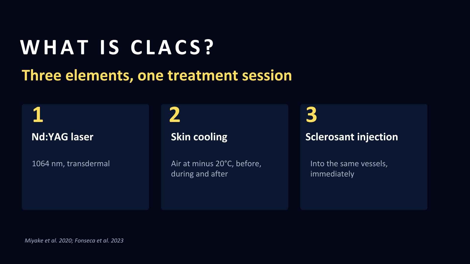
CLaCS combines three elements delivered in one treatment session: transdermal 1064 nm Nd:YAG laser, skin cooling with air at minus 20 degrees Celsius blown onto the skin and needle before, during and after treatment, and immediate injection of a sclerosant into the same vessels [3,4].
The sclerosant in the original technique is not a detergent. Miyake and colleagues described 75% dextrose, an osmotic agent, chosen precisely because the laser has already reduced vessel calibre and a less aggressive agent is therefore sufficient [4]. This matters more than it first appears, and I will return to it.
The rationale is that laser exposure produces endothelial injury, vasospasm and reduction in vessel calibre before injection, which may allow a lower sclerosant volume and a less aggressive inflammatory response than injection alone [1,4]. Cooling provides analgesia and some epidermal protection. Augmented reality vein imaging is often used alongside it to map reticular feeders.
That rationale is plausible. It is a reason to test the technique, not evidence that the technique works better.
The original technical description, reporting preliminary outcomes in lower limb reticular veins, gave satisfactory vessel elimination in 86% of cases, with ecchymosis in up to 30% and intravascular thrombus in 14% [4]. Those figures are not better than Microsclerotherapy performed well, and they were never intended as comparative data.
3. The 2026 Meta-analysis: What It Pooled
The new synthesis searched PubMed, Embase and CENTRAL to February 2025 and included six studies covering 813 patients or treated limbs [1].
Two things about that description deserve attention before any of the results.
First, the design mix. Six studies comprise three randomised controlled trials, one prospective non-randomised comparative study and two retrospective single-arm cohorts [1]. Only three datasets were directly comparative, so every pooled effect estimate in this review rests on three studies, one of which is not randomised.
Second, the search date. The literature search ran to February 2025, and the paper is dated 2026. There is a gap of roughly eighteen months between the last search and publication, which is long enough to matter in an area of research where individual trials arrive at the rate of about one a year.
The review itself reports some concerns or higher risk of bias in multiple included studies, substantial heterogeneity for several endpoints, and no ability to assess publication bias because no outcome had ten or more studies [1]. Those are the authors' own statements, and they are appropriately candid.
A note on evidence grading. Pooled data from a meta-analysis is sometimes labelled level 1a as a matter of course. I do not think that label survives the scrutiny of this review. A pooled estimate drawn from three studies, one non-randomised, with acknowledged risk-of-bias concerns and high heterogeneity on some endpoints, is evidence from meta-analysis, but with obvious reasons to downgrade our confidence in it. It is better than what we had. It is not the same thing as a large, low-bias, replicated randomised comparison, and describing it that way would mislead anyone making a purchasing decision on the strength of it.
4. Does CLaCS Reduce Matting, Microthrombi, and Ecchymosis?
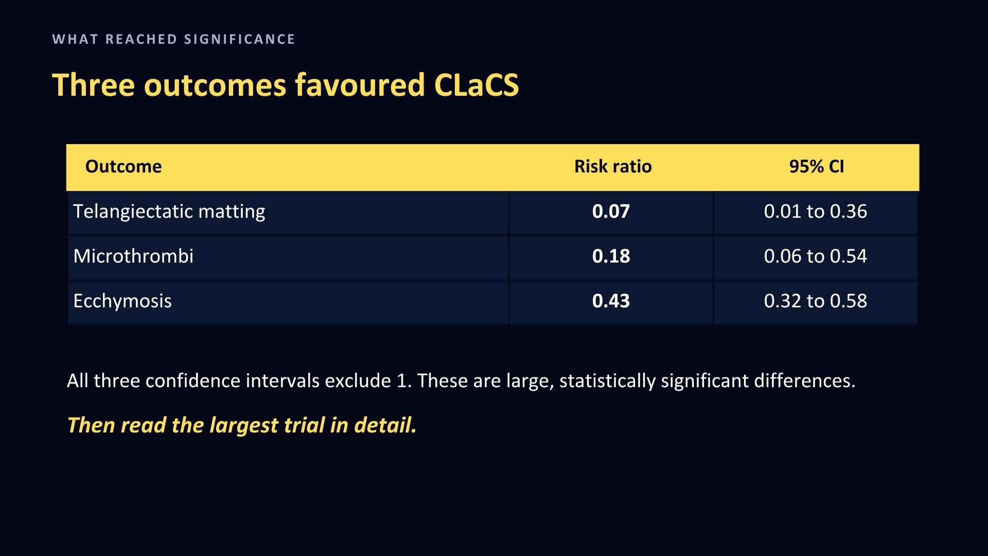
Against sclerotherapy alone, the meta-analysis reported [1]:
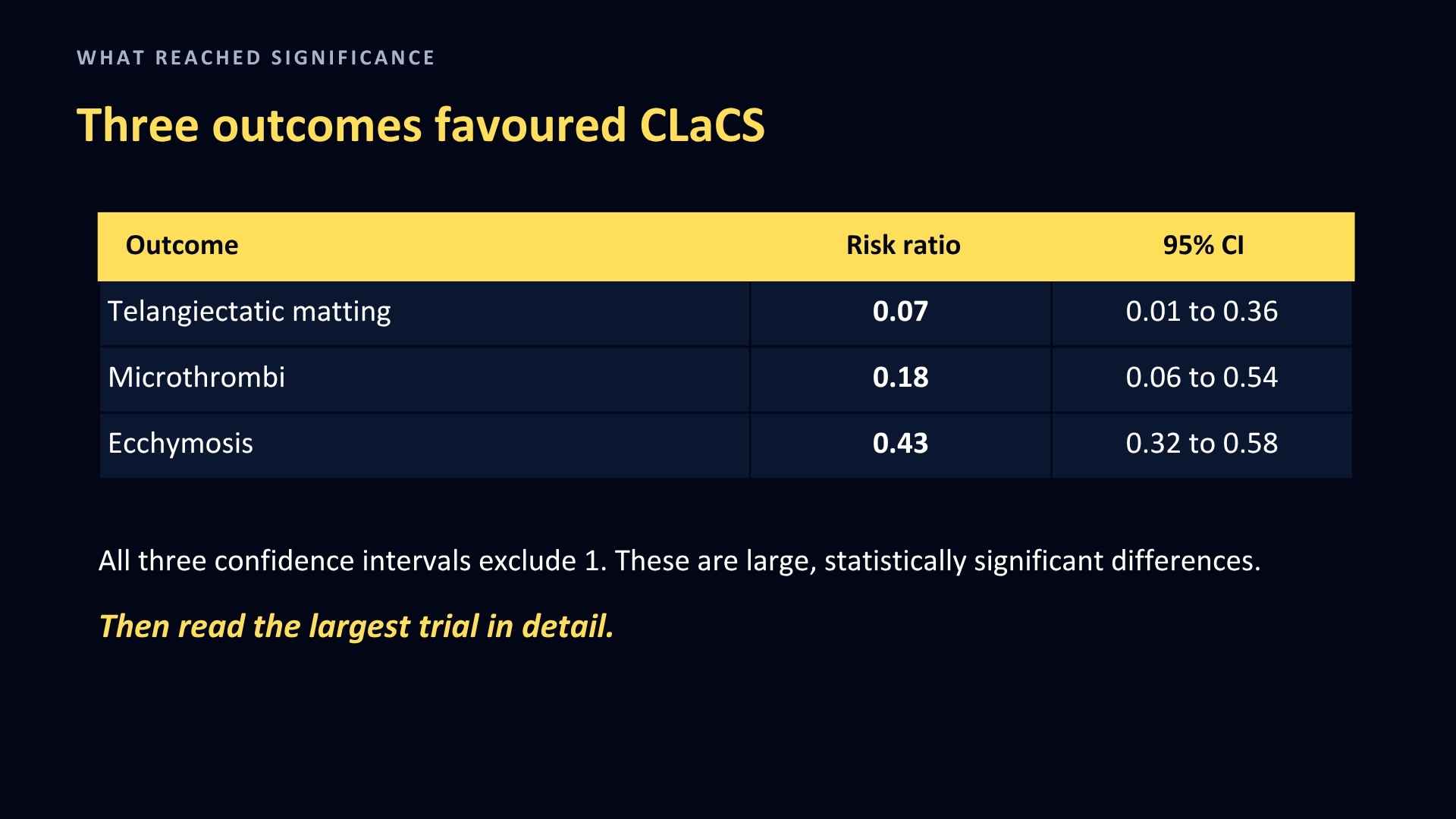
A risk ratio below 1 means fewer events with CLaCS, and in all three cases the confidence interval does not cross 1, so these are statistically significant reductions rather than chance findings. They are also large differences. Taken at face value, that is a clinically significant result about three problems patients notice and complain about, and matting is the one that most reliably turns a technically sound treatment into an unhappy patient.
But when you read the publication of the largest trial in detail, these differences may not be so compelling.
What the comparator arm received
Nasser and colleagues randomised 400 patients at Cairo University Hospitals, 200 to each treatment arm, with 195 and 197 analysed after eight were lost to follow-up [2]. This single trial supplies most of the events behind the pooled estimates, so its methods deserve close reading.
Group A received Nd:YAG 1064 nm laser at 50 to 70 J per square centimetre with continuous air cooling, followed by 12 to 15 mL of 70% dextrose through a 30 gauge needle. No compression afterwards, and a hirudin-based gel twice daily for a week.
Group B received 8 to 10 mL of 0.5% polidocanol foam, prepared by the Tessari method, into the same 20 cm by 20 cm field, followed by compression stockings for a week [2].
That is the comparison. Not CLaCS against Microsclerotherapy. CLaCS against a large volume of 0.5% polidocanol foam delivered into a single 400 square centimetre area of cosmetic C1 disease.
Why that matters so much
The complication rates in Group B tell you what that protocol did to those patients. Pigmentation was assessed at eight weeks after completion of the treatment course, and complications were recorded across the study [2]:
A moderate pigmentation rate of 77.7% is a striking figure. For an internal comparison within this same article, the sclerotherapy control arm in the Diken trial, using liquid polidocanol at 0.5% to 1.0%, had a hyperpigmentation rate of 28.5% [8]. A matting rate of 19.3% and a skin necrosis rate of 4.2% in cosmetic C1 patients are also high for a treatment sought purely for appearance.
In fairness to the trial, pigmentation in that arm did settle substantially with time: over a six-month follow-up period the rate fell from 77.7% to 45% [2]. The authors themselves suggest the eight-week figure partly reflects how early patients were reviewed. That is a reasonable point, and it should be stated alongside the headline number. A 45% pigmentation rate at six months is still high.
So what the largest trial in this meta-analysis actually shows is that CLaCS was compared against a sclerotherapy protocol that performed poorly, and it is not possible to tell from these data how much of the difference reflects the laser and how much reflects foam at 0.5% in 8 to 10 mL volumes with no evident concentration titration.
The trial authors are open about part of this. They attribute the lower complication rate in the CLaCS arm to the milder effects of dextrose compared with polidocanol [2]. What they do not address is whether a detergent foam at that concentration and volume was the right comparator in the first place.
The consequence for the pooled estimates
Three specific consequences follow.
The matting risk ratio of 0.07 rests on a trial in which one group had no events at all: no matting in 195 CLaCS patients, against 38 cases in 197 sclerotherapy patients. When one treatment arm records zero events, the ratio cannot be calculated precisely, and that is why the confidence interval is so wide, running from 0.01 to 0.36. A figure of 0.07 should therefore be read as "much less matting in this trial" and not as a treatment effect size to quote to a patient.
The treatment arms also differed in aftercare, not only in modality and sclerosant. Group A had no compression and a topical antithrombotic gel; Group B had a week of compression stockings. Retained thrombus is an important driver of staining, and the two treatment arms managed it differently.
And the comparison is between a hybrid technique and a specific sclerotherapy protocol, not between a hybrid technique and best-practice Microsclerotherapy. Those are different questions, and only the first has been answered.
Other things worth knowing about this trial
Every participant was female, in both treatment arms. That was not an eligibility criterion: the authors attribute it to the fact that those presenting for aesthetic treatment of C1 disease were women, and they list it among their own limitations [2]. The trial restricted entry to vessels under 1.5 mm in the standing position and Fitzpatrick types II to IV. The primary assessment was at 8 weeks after each session, with pigmentation followed to six months. Duplex was used to exclude anyone with superficial or deep venous reflux, which is a real strength and makes this a cleaner C1 population than most.
Two reporting inconsistencies are worth noting for anyone reading the paper. The patient flow figure does not reconcile: 457 assessed, an exclusion box reading 13 and 57, and 400 randomised. The severe pigmentation figure for Group A is printed as 0.05% where 1 of 195 is 0.5%. Neither error changes the direction of the findings, but both are worth noticing in the trial carrying most of the weight in the pooled analysis.
One claim in the discussion needs correcting. The authors state that CLaCS has been incorporated into the ESVS clinical practice guidelines on lower limb chronic venous disease [2]. Having now read the guideline text, that is not what it says.
CLaCS appears in the body text of the ESVS 2022 guidelines exactly once, in section 4.5.3 on hybrid techniques, in a single sentence describing it as a new option for treating telangiectasias, reticular and feeder veins with promising results, while stating that more studies are required for its validation [6].
That is a mention with an explicit caveat about validation, not an incorporation. CLaCS has no numbered recommendation, no class, and no level of evidence anywhere in the document. To describe it as incorporated into the guidelines, without quoting the qualification attached to it in the same sentence, overstates the position.
This is not a quibble about wording. The overstatement here appears in a peer-reviewed randomised trial in a major vascular journal, which is precisely where a busy clinician would expect to be able to take a guideline claim at face value. If it can pass peer review there, it can certainly travel into a course brochure or a device brochure, and by then the validation caveat will have been lost entirely. When a technique is described to you as being in the guidelines, the question to ask is which recommendation number, what class, and what level.
5. Does CLaCS Reduce Post-Sclerotherapy Pigmentation?
A note on terminology. Many of the trials in this area of research record their outcome as hyperpigmentation, and where I report what a trial measured I have kept its word. The better term for what we are discussing is post-sclerotherapy pigmentation. Hyperpigmentation strictly means excess melanin production, whereas the discolouration seen after sclerotherapy is a combination of melanin and haemosiderin. That distinction is not pedantry here: it is the reason the results in this section are harder to interpret than they first appear, because a trial measuring colour change is capturing two different processes with different time courses and different responses to treatment.
If CLaCS produces fewer microthrombi, and retained intravascular blood is a recognised precursor of haemosiderin staining, then CLaCS should produce less pigmentation. The meta-analysis makes exactly that argument, and it is a reasonable one [1].
The data do not support this argument. The random-effects pooled estimate for hyperpigmentation was not statistically significant and heterogeneity was very high [1]. The result depends heavily on how pigmentation was measured, with photographic and colorimetric assessment giving different signals.
The within-patient randomised trial by Rodrigues and colleagues shows what this looks like at study level [5]. Reticular veins were treated in each patient with CLaCS on one limb and 0.5% polidocanol foam on the other, with blinded evaluators, at 60 days after a single session. There was no significant difference in the number of limbs developing pigmentation, either by photographic assessment (7 CLaCS against 5 foam) or by colorimetry (9 against 13). The CLaCS side did show significantly lower pigmentation intensity on colorimetry, with a mean delta E of 1.30 against 1.44 [5].
That last figure needs interpreting rather than reporting. A delta E difference of 0.14, with both values close to the threshold at which a colour difference becomes perceptible to the human eye at all, is a statistically significant difference of doubtful visible consequence. That is my interpretation and not the authors' conclusion, and colorimetric thresholds are not settled. But a patient asking "will I stain less?" is asking about what she can see in the mirror, and this trial does not clearly answer yes.
The same trial did find less microthrombus formation and drainage, less bruising, and a lower volume of sclerosant needed, with no difference in vein diameter reduction and no major adverse events [5]. That is consistent with the pooled picture: the short-term procedural experience looks better, the pigmentation outcome does not clearly follow, and the clearance outcome is flat.
CLaCS has not been shown to reduce pigmentation, despite a good biological reason to expect that it should. That is an unusual and interesting result, and it is the part of this evidence base I would most like to see settled.
6. Is CLaCS More Effective for Leg Vein Clearance Than Sclerotherapy?
The review did not produce a pooled comparative estimate for vessel clearance. The included studies defined and measured clearance in ways too different to combine, so the authors described their findings one by one instead of calculating a single figure [1].
This is the single most important limitation of the evidence. Clearance is the outcome the patient is paying for. Individual studies point in different directions: Nasser reported markedly better complete elimination with CLaCS across three sessions [2], while Rodrigues found no difference in diameter reduction between limbs [5]. Those are not reconcilable into a single number, and the review was right not to try.
The consequence is that there is currently no pooled evidence that CLaCS clears leg veins better than optimised Microsclerotherapy. What the evidence suggests is that it may clear them about as well, with fewer visible side effects in the short term. Those are different claims and they should not be blurred together in a patient consultation.
7. Where This Evidence Comes From
The published comparative primary evidence comes mainly from Brazil and Egypt [2,3,5], with a further comparative study from Russia by Rosukhovskiy, published in Flebologiya in 2021, which compares CLaCS with Microsclerotherapy [12]. That paper is not indexed on PubMed and I have not read it in full, so I cannot comment on its methods. The meta-analysis states that CLaCS has been adopted in more than 30 countries, and that is an author-reported contextual statement rather than verified utilisation data [1]. International diffusion is real; global mainstream adoption is not established.
On declared interests and the independence of the evidence base, the meta-analysis declares no conflicts of interest and no specific funding [1]. It also broadens authorship compared with the earlier CLaCS literature. Even so, the underlying evidence remains concentrated in a relatively small research community, and there is direct author overlap between the meta-analysis and at least one of its included primary trials [1,5].
Let me be clear about what I am saying. I am not alleging any misconduct. Author overlap between a trial and a subsequent synthesis is common and often unavoidable in a very specialised area of research. I am making a comment about the relative lack of independent replication, and independent replication is what should determine our confidence in any new technique. At present, a clinician relying on this evidence is relying largely on a single investigator network. That is a reason for measured adoption, not for suspicion, and it is exactly the gap that a trial run by a different group in a different country would close.
8. The Comparison That Matters for UK Clinics
For most UK practitioners the real question is not "is CLaCS better than injecting". It is whether CLaCS is the treatment option worth spending a substantial equipment budget on.
CLaCS requires three pieces of equipment rather than one: an Nd:YAG platform, a cold-air skin cooling system, and an infrared vein imaging device, plus competence in both laser and injection treatment [1,4]. In the Nasser protocol that meant an Alma Lasers Nd:YAG, a Zimmer cooler and a VeinViewer Flex [2]. No health economic analysis has been published, and the meta-analysis offers none [1]. Reducing visible short-term sequelae is clinically worthwhile, but it is not the same as cost-effectiveness.
The obvious alternative, for a substantially smaller budget, is 4 MHz radiofrequency thermocoagulation used alongside Microsclerotherapy. This is what we teach, so read what follows with that in mind.
The supporting evidence. Diken and colleagues randomised 111 patients with CEAP C1 spider veins to liquid sclerotherapy alone or sclerotherapy immediately followed by low-energy percutaneous radiofrequency thermocoagulation, using a TC3000 generator at 20% power with 0.2 second pulses and 0.075 mm Ballet microneedles [8]. The trial itself does not state an operating frequency. The FDA 510(k) summary for the Veinwave/TC3000 records that the system uses a current of 4 MHz, delivered monopolar at approximately 30 W, for treating lower limb spider veins and telangiectasia by thermocoagulation, and lists the Ellman Surgi-Max, also 4 MHz, as a predicate [10]. One caveat on that identification: Diken attributes the TC3000 to F Care Systems of Belgium, while the 510(k) was submitted by Newlands Clinical Trials of Bolton. The trade name and model number match, and these devices have moved between distributors over the years, but I have not been able to confirm from either document that they are the same generator. On the balance of the evidence Diken tested at 4 MHz, and that is how I read it, but it is an inference rather than a statement in the trial.
One detail is worth noting for anyone reproducing the protocol. The 510(k) summary describes power settings between 30% and 60% in 5% increments [10], while Diken deliberately worked at 20%, having found in preliminary work that 10% produced no effect and that 30% and above is used for ablation rather than spasm [8]. Their intention was to provoke venospasm without injuring the vein wall. Whether that is what actually happened is something the authors are careful to say they cannot confirm without histology.
The result is more specific than a summary suggests, and the specificity is the useful part. Venous clinical severity scores fell significantly in both treatment arms, and the type of intervention made no difference to that fall. What did differ was hyperpigmentation (28.5% against 9%), trapped blood (16% against 3%), self-assessed cosmetic satisfaction, and the number of sessions needed (3.3 against 2.7). Skin necrosis did not differ [8].
Three further details matter. Both treatment arms received identical compression, 36 hours of elastic bandaging followed by class 1 stockings for three months, so unlike the Nasser trial the aftercare was not a confounder. Patients with axial reflux and those with feeder veins larger than 1 mm were excluded, and only one spider vein was targeted per session. And the combined procedure took considerably longer, 25.5 minutes against 7.2 minutes [8].
So the combination did not treat the veins better. It produced fewer of the things patients dislike about the treatment, in fewer visits but longer visits. That is a narrower claim than "improves outcomes" and it is the claim I would defend.
The limitations are equally worth stating. This was a single-centre pilot with 56 and 55 patients per arm, the cosmetic outcome was self-assessed on an unvalidated three-point scale, and the authors are explicit that they do not know whether the radiofrequency produced vein spasm or vein injury [8].
What the 2026 TYPER trial adds. TYPER randomised 41 patients, 20 analysed per arm, to transdermal radiofrequency plus polidocanol sclerotherapy or polidocanol sclerotherapy alone, in a single session with assessment at 30 days [9].
The concentrations deserve attention, because they are the ones this article has been arguing for throughout: liquid polidocanol at 0.25% for telangiectasias and 0.5% for reticular veins, both within the European guideline range and at its lower end [9]. This is the only trial discussed here that used a comparator most of us would recognise as ordinary good practice.
Both treatment arms improved significantly and quality of life improved in both. The authors concluded that transdermal radiofrequency was not superior to standard care.
This is the most important finding for anyone weighing the combination. TYPER measured hyperpigmentation directly and found it identical at 65% in both treatment arms, while the radiofrequency arm developed crusting that the sclerotherapy arm did not. That is a negative result on precisely the endpoint where Diken reported a benefit.
Why this does not simply overturn the Diken finding. Two differences between the designs of the trials are large enough that the findings of one cannot simply be applied to the other, and I set them out so readers can judge for themselves rather than take my word for it.
The first is the device and the frequency. TYPER used the V-Eraser, whose specification gives a fundamental frequency of 450 kHz and maximum output of 19.5 W [11]. Diken used the Veinwave/TC3000 at 4 MHz and approximately 30 W [10]. Operating frequency affects how energy is deposited in tissue, so a null result at 450 kHz should not be assumed to apply at 4 MHz. No study has compared the two frequencies directly, so this is a reason to hold back from that assumption rather than evidence that 4 MHz performs better.
The second difference is the technique itself, and it is arguably the bigger one. TYPER delivered sclerotherapy and radiofrequency simultaneously, with the energy passing through the needle tip inside the vessel lumen. Diken performed sclerotherapy first, then applied a separate 0.075 mm micro-needle perpendicular to the skin over the vein, without a second puncture, aiming to provoke venospasm rather than to ablate [8,9]. The TYPER paper itself describes these as the two distinct ways transdermal radiofrequency can be delivered [9]. They are different procedures that happen to share a category name.
Where that leaves the evidence for what we teach. One randomised pilot at 4 MHz, sequential technique, showing less pigmentation and less trapped blood but no difference in venous clinical severity score. One randomised trial at 450 kHz, simultaneous technique, showing no clearance benefit, no pigmentation benefit, and some added crusting. No trial has replicated the Diken result, and no trial has compared the two techniques head to head.
The TYPER authors report that the Brazilian Society of Angiology and Vascular Surgery classifies transdermal radiofrequency for CEAP C1 as recommendation IIa, level of evidence C, and states explicitly that randomised trials are still required [9]. I have not read that guideline at source, so this is reported at second hand. Level C is expert opinion. That is a fair summary of where this sits, and it is a weaker position than the one I would have described a year ago.
One hybrid the ESVS describes more favourably. Worth noting, because it is adjacent to CLaCS and often confused with it: the ESVS guidelines state that 1064 nm Nd:YAG long pulse laser applied after foam sclerotherapy with polidocanol has proven more effective than sclerotherapy alone at three months, two years and three years [6]. That is laser after injection, rather than laser before it, and it carries longer follow-up than anything in the CLaCS literature. It still attracts no separate graded recommendation, but the evidence behind it is described in stronger terms than the CLaCS sentence.
The comparison nobody has made. There has never been a head-to-head trial of CLaCS against 4 MHz thermocoagulation combined with Microsclerotherapy. Both approaches have evidence pointing the same way: better short-term cosmetic and adverse-event outcomes, no demonstrated clearance advantage. One requires an Nd:YAG platform, cooling and imaging; the other requires a generator costing a small fraction of that. Anyone who tells you which is clinically better is giving you an opinion.
What I would say to a clinician deciding. Look again at those two comparator arms before you look at any equipment. Nasser's sclerotherapy patients received 0.5% polidocanol foam in 8 to 10 mL volumes and 77.7% of them developed moderate pigmentation. Diken's sclerotherapy patients received liquid polidocanol at 0.5% to 1.0% and 28.5% of them developed hyperpigmentation. Both sit above the European guideline range for telangiectasia, which is 0.25% to 0.5% for polidocanol, with 0.5% to 1.0% reserved for reticular veins. TYPER, which used 0.25% and 0.5% correctly, found that adding energy to a properly dosed injection produced no significant advantage on any outcome it measured [9].
The most reliable route to less matting, less trapped blood and less staining is not a machine. It is the lowest effective concentration, accurate superficial placement, small volumes per site, review at one to two weeks to evacuate retained coagulum, and firm advice to the patient to keep the treated area out of the sun while it settles. Ultraviolet exposure during the inflammatory phase drives the melanin component of post-inflammatory pigmentation, and it costs nothing to prevent.
Buy equipment to extend the range of vessels you can treat, particularly at the fine end where injection is unreliable. Do not buy it to compensate for a protocol that could be corrected on Monday morning.
9. When It Might Be Appropriate to Consider CLaCS
Where it may have a place [1,4]:
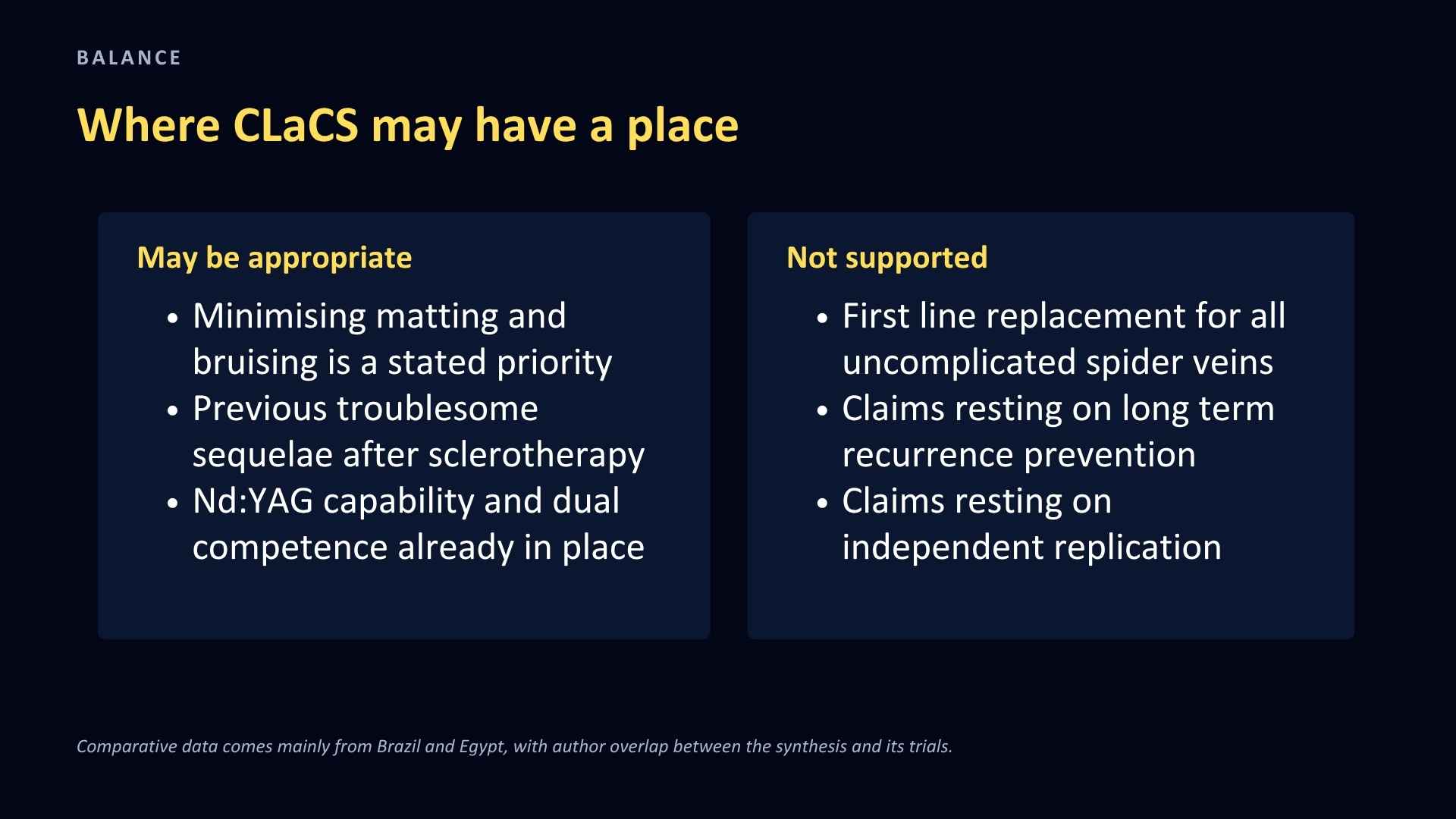
* Specialist venous or aesthetic practice where minimising matting, microthrombi and ecchymosis is a stated priority for the patient
* Patients with a history of cosmetically troublesome sequelae after standard sclerotherapy, where reducing short-term visible adverse events is the main treatment goal
* Settings that already have Nd:YAG capability, cooling, and operators competent in both laser and injection
* Selected patients where an experienced operator judges the combination useful, provided the consent conversation states plainly that broad superiority is unproven
Not supported as routine [1,6,7]:
* First-line replacement for all uncomplicated leg spider veins. Broad superiority over optimised Microsclerotherapy is not established
* Settings without specialist laser expertise, adequate cooling, or the ability to counsel honestly about the uncertainty
* Cases where the goal is straightforward low-cost clearance and the patient accepts the known adverse-event profile of Microsclerotherapy
* Any claim that depends on long-term recurrence prevention, standardised clearance benefit, or independent replication, because none of those exists yet
On the guidelines. The ESVS 2022 position is worth setting out precisely, because it is the guideline most often invoked in this discussion [6]:
* Recommendation 40 (Class I, Level A): for patients with reticular veins where treatment is planned, sclerotherapy is recommended as the first choice treatment.
* Recommendation 41 (Class IIa, Level A): for patients with telangiectasias where treatment is planned, sclerotherapy should be considered.
* Recommendation 42 (Class IIa, Level B): for patients with telangiectasias where treatment is planned, transcutaneous laser should be considered.
* CLaCS: not the subject of any recommendation. It appears in the body text exactly once, in a single descriptive sentence in section 4.5.3 on hybrid techniques, which calls it a new option for treating telangiectasias, reticular and feeder veins with promising results, and states that more studies are required for its validation. Because it is not the subject of a recommendation, no class of recommendation and no level of evidence are attached to it.
So the strongest graded recommendation in this area belongs to sclerotherapy, and CLaCS sits outside the recommendation structure altogether. That is not a criticism of the technique. It is where a new modality sits before the trials that would grade it have been done.
Being mentioned in a guideline and being recommended by one are different things, and as section 4 sets out, that difference has already been blurred once in the published literature. Look for the numbered recommendation and its class and level, not the mention.
10. Duplex Ultrasound: Unchanged by Any of This
None of this new evidence alters duplex policy, and the source material is right to keep the two questions separate.
Duplex remains indicated where there are venous symptoms, visible varicose veins, oedema, skin changes, a history of ulceration or deep vein thrombosis, previous venous intervention, or telangiectasia resistant to previous treatment. It is not routinely required for isolated, asymptomatic, uncomplicated thread veins in the absence of those features, a position taken by both the UIP Microsclerotherapy guidance and the 2023 SVS/AVF/AVLS guidelines [7,13]. The ESVS 2022 guidelines take a more conservative line: Recommendation 38 states that duplex should be performed before treating all patients presenting with reticular veins or telangiectasias, to look for associated incompetent veins. It is Class I but Level C, meaning expert consensus rather than trial data [6]. I set out the reasoning for the selective position in an earlier article, and nothing in the CLaCS literature changes it.
It is worth noting that the Nasser trial excluded anyone with duplex-confirmed reflux [2]. Its findings therefore apply to an uncomplicated C1 population, which is a strength of that trial rather than a limitation.
11. Key Clinical Points
* The 2026 meta-analysis is the first pooled comparison of CLaCS against sclerotherapy alone: six studies, 813 patients or limbs, but only three directly comparative datasets, one of them non-randomised [1].
* Pooled reductions were reported for telangiectatic matting (RR 0.07, 95% CI 0.01 to 0.36), microthrombi (RR 0.18, 0.06 to 0.54) and ecchymosis (RR 0.43, 0.32 to 0.58) [1].
* Those results are best described as evidence from meta-analysis, but with obvious reasons to downgrade our confidence in it: risk of bias, heterogeneity and sparse events. They are not clean level 1a evidence.
* Post-sclerotherapy pigmentation was not significantly reduced on random-effects pooling, with very high heterogeneity, despite a good biological reason to expect that it would be [1,5].
* Vessel clearance could not be pooled at all because of heterogeneous definitions. There is no pooled evidence that CLaCS clears leg veins better than Microsclerotherapy [1].
* The largest comparative trial compared CLaCS not against Microsclerotherapy but against 8 to 10 mL of 0.5% polidocanol foam delivered into a 20 cm by 20 cm field, a protocol that produced moderate pigmentation in 77.7% of patients and matting in 19.3% [2].
* The modality, the sclerosant and the aftercare all differed between those arms, so the laser effect cannot be separated from the effect of comparing against a harsh sclerotherapy protocol [2].
* The pooled matting estimate rests on a trial in which one treatment arm recorded no events at all (0 of 195 against 38 of 197), which is why its confidence interval is so wide [1,2].
* The comparative evidence remains geographically and institutionally concentrated, with author overlap between the synthesis and its included trials [1,5].
* The ESVS 2022 guidelines give sclerotherapy a Class I Level A recommendation as first choice for reticular veins and Class IIa Level A for telangiectasias. CLaCS is not the subject of any recommendation, appearing only in a single descriptive sentence stating that more studies are required for its validation [6].
* No head-to-head trial compares CLaCS with 4 MHz radiofrequency thermocoagulation combined with Microsclerotherapy.
* Randomised evidence at 4 MHz shows less pigmentation, less trapped blood and fewer sessions with adjuvant thermocoagulation, but no difference in venous clinical severity score [8]. The device used, the Veinwave/TC3000, is confirmed at 4 MHz in its FDA 510(k) summary [10]. The 2026 TYPER trial found no clearance benefit, but used a 450 kHz device [9,11], so its result does not transfer to 4 MHz equipment.
* Concentration discipline remains the most reliable intervention available: European guideline ranges are 0.25% to 0.5% polidocanol for telangiectasia and 0.5% to 1.0% for reticular veins. The Nasser and Diken comparator arms sat at or above the top of those ranges [2,8]. The one trial that used guideline-appropriate low concentrations, TYPER, found that adding radiofrequency produced no clearance and no pigmentation benefit [9].
* Sun avoidance during the inflammatory phase, alongside review at one to two weeks to evacuate retained coagulum, addresses pigmentation at no capital cost.
* CLaCS is a reasonable specialist option where minimising short-term visible sequelae is the priority. It is not a demonstrated replacement for well-performed Microsclerotherapy.
12. Frequently Asked Questions (FAQ)
Q: What does the 2026 meta-analysis show about CLaCS for leg spider veins? A: The 2026 systematic review and meta-analysis shows CLaCS achieves statistically significant reductions in telangiectatic matting, microthrombi, and ecchymosis compared to isolated sclerotherapy [1]. However, it found no statistically significant reduction in post-sclerotherapy pigmentation, and vessel clearance could not be pooled due to high heterogeneity across studies [1].
Q: Is CLaCS officially recommended in the ESVS Clinical Practice Guidelines? A: No. CLaCS is mentioned once in the body text of the ESVS 2022 Guidelines (Section 4.5.3) as a novel hybrid technique with promising results requiring further validation [6]. It carries no formal recommendation number, class of recommendation, or level of evidence [6]. Sclerotherapy remains the Class I, Level A recommended treatment for reticular veins [6].
Q: Does CLaCS clear leg veins better than standard microsclerotherapy? A: There is currently no pooled comparative evidence proving CLaCS provides superior vessel clearance compared to well-performed microsclerotherapy [1]. Clearance definitions varied widely across included trials, preventing direct statistical synthesis [1].
13. References
1. Simoni GH, Madera D, Papatheodorou N, Neves S, Varella ACP, Alonso JG, Mahfouz N. Cryo-laser cryo-sclerotherapy versus isolated sclerotherapy for telangiectasias and reticular veins: a systematic review and meta-analysis. J Vasc Surg Venous Lymphat Disord. 2026. doi:10.1016/j.jvsv.2026.102582. Accepted journal pre-proof. No PMID available at the time of writing; citation details should be confirmed against the final published version. https://www.jvsvenous.org/article/S2213-333X(26)00142-3/fulltext
2. Nasser MM, Ghoneim BM, Eldaly W, Elmahdy H. A comparative study between cryo-laser cryo-sclerotherapy and sclerotherapy in the treatment of telangiectasia and reticular veins: a randomized controlled trial. J Vasc Surg Venous Lymphat Disord. 2024;12(4):101874. PMID: 38522666. https://pubmed.ncbi.nlm.nih.gov/38522666/
3. Fonseca MM, Mocelin FJ, Grill MH, Gianesini S, Miyake K, Argenta R, Pereira AH. Nd:YAG laser combined with injection sclerotherapy in the treatment of reticular veins and telangiectasias (CLaCS method): a triple-blind randomized clinical trial comparing two sclerosing agents associated with same laser patterns. Phlebology. 2023;38(3):165-171. PMID: 36657386. https://pubmed.ncbi.nlm.nih.gov/36657386/
4. Miyake RK, Chi YW, Franklin IJ, Gianesini S. State of the art on cryo-laser cryo-sclerotherapy in lower limb venous aesthetic treatment. J Vasc Surg Venous Lymphat Disord. 2020;8(5):893-895. PMID: 32179040. https://pubmed.ncbi.nlm.nih.gov/32179040/
5. Rodrigues A, Puggina J, da Silva VS, Ribeiro CM, Santiago FR, Neves S, Simoni GH, Portugal MF, Ramacciotti E. Pigmentation after foam or cryo-laser cryo-sclerotherapy for lower limb reticular veins: a within-patient randomized trial. J Vasc Surg Venous Lymphat Disord. 2025;13(6):102285. PMID: 40653087. https://pubmed.ncbi.nlm.nih.gov/40653087/
6. De Maeseneer MG, Kakkos SK, Aherne T, et al. Editor's Choice: European Society for Vascular Surgery (ESVS) 2022 Clinical Practice Guidelines on the Management of Chronic Venous Disease of the Lower Limbs. Eur J Vasc Endovasc Surg. 2022;63(2):184-267. PMID: 35027279. https://pubmed.ncbi.nlm.nih.gov/35027279/
7. Gloviczki P, Lawrence PF, Wasan SM, et al. The 2023 Society for Vascular Surgery, American Venous Forum, and American Vein and Lymphatic Society clinical practice guidelines for the management of varicose veins of the lower extremities. Part II. J Vasc Surg Venous Lymphat Disord. 2024;12(1):101670. PMID: 37652254. https://pubmed.ncbi.nlm.nih.gov/37652254/
8. Diken Aİ, Alemdaroğlu U, Özyalçın S, Hafez İ, Tünel HA, Yalçınkaya A, Ecevit AN. Adjuvant radiofrequency thermocoagulation improves the outcome of liquid sclerotherapy in the treatment of spider veins of the leg: a pilot study. Phlebology. 2021;36(8):620-626. PMID: 33813962. https://pubmed.ncbi.nlm.nih.gov/33813962/
9. Higino Becker R, Caffaro RA, de Oliveira CCC, Marcondes RP, Negrão AR, Magella F, Volpiani GG, Neves S, Rodrigues RCB, Rodrigues MFBF, da Silva VS, Ribeiro CM, Kikuchi R, Santiago FR, Ramacciotti E. Transdermal radiofrequency (V Eraser) plus polidocanol sclerotherapy versus polidocanol sclerotherapy alone for the treatment of reticular veins and telangiectasias: a randomized controlled trial (TYPER trial). Phlebology. 2026;41(5):410-418. PMID: 40905659. https://pubmed.ncbi.nlm.nih.gov/40905659/
10. US Food and Drug Administration. 510(k) Premarket Notification Summary, Veinwave/TC3000. 510(k) Number K083352. Newlands Clinical Trials Ltd. Substantial equivalence determination issued 12 June 2009. Records monopolar operation at 4 MHz, approximately 30 W balanced at 500 ohms, for epilation and treatment of lower limb spider vein or telangiectasia by thermocoagulation. Regulatory submission document. Cited for device operating frequency and intended use.
11. V Company do Brasil. V-Eraser: technical specifications. Maximum output power 19.5 W, fundamental frequency 450 kHz. http://vcompanydobrasil.com/v-eraser-v-01/ Manufacturer specification, not peer-reviewed. Cited only for the device operating frequency.
12. Rosukhovskiy DA. Comparative study of cryo-laser and cryo-sclerotherapy with microsclerotherapy. Flebologiya. 2021;15(3). Not indexed on PubMed. Cited only as the source of the Russian comparative data referred to in reference 1; not read in full and not relied on for any claim in this article.
13. Tan M, Shaydakov E, Parsi K, Davies AH; on behalf of the International Union of Phlebology. Microsclerotherapy: International Union of Phlebology One-Page Guidelines. Phlebology. 2024;39(4):280-283. PMID: 38103047. https://pubmed.ncbi.nlm.nih.gov/38103047/
About the Author

This educational article is written and regularly reviewed by Dr Haroun Gajraj, a GMC-registered vein specialist who has treated thousands of patients with vein disease and has trained many doctors and nurses in microsclerotherapy, radiofrequency thermocoagulation, and related cosmetic vein procedures. Dr Gajraj is the founder and board member of the British Association of Sclerotherapists. You can view his current GMC registration and independent patient reviews on iWantGreatCare for further information about his clinical background.
Clinicians remain responsible for assessing each patient, obtaining informed consent, explaining risks and alternatives, and working within the scope of their professional registration and regulatory guidance.
Subscribe
For regular updates get my weekly newsletter here
© VeinCare Academy | Dr. Haroun Gajraj | veincare.academy
This article is intended for qualified healthcare professionals. All clinical decisions should be based on individual patient assessment, primary medical literature and current professional guidelines.