- May 20
Brown Marks After Microsclerotherapy:Causes, Prevention and Treatment.
- Haroun Gajraj
- 0 comments
By Dr. Haroun Gajraj | VeinCare Academy | 20th May 2026
This article is written for healthcare professionals who perform microsclerotherapy for leg spider veins and reticular veins. It covers the incidence, pathological mechanisms, risk factors, evidence-based prevention strategies, and treatment options for post-sclerotherapy hyperpigmentation (PSH) — the most common cosmetically significant complication of spider vein treatment.
🎥 Prefer video? Watch the full 12-minute clinical guide on YouTube: "Brown Marks After Microsclerotherapy — What Every Practitioner Needs to Know" →
Contents
1. What is post-sclerotherapy hyperpigmentation?
2. How common is it? — Incidence
3. What causes brown marks? — The dual pigment mechanism
4. Two clinical patterns
5. Who is at risk? — Risk factors
6. How to prevent post-sclerotherapy pigmentation — 6 evidence-based strategies
7. How to treat established pigmentation
8. Management algorithm
9. Key takeaways
10. References
1. What Is Post-Sclerotherapy Hyperpigmentation?
Post-sclerotherapy hyperpigmentation (PSH) refers to brown or brownish-black discolouration of the skin that develops following sclerotherapy, typically tracking the course of treated vessels. It is the most commonly reported cosmetically significant adverse sequela of microsclerotherapy for leg spider veins and reticular veins [1][5][23].
For patients who have chosen sclerotherapy specifically to improve the appearance of their legs, brown marks can be more distressing than the original spider veins — making this complication particularly damaging to patient satisfaction and the practitioner-patient relationship. The good news is that PSH is largely preventable when the underlying mechanisms are understood and appropriate clinical protocols are followed [5][23].
2. How Common Is It? — Incidence
The published incidence of PSH varies widely, primarily because it is so strongly influenced by sclerosant concentration and technique. The most frequently cited range — 10 to 30% of patients short-term — comes from the landmark 1995 paper by Goldman, Sadick and Weiss in Dermatologic Surgery [5], which established the benchmark for spider and reticular vein treatment across vessels of 0.1–5 mm diameter.
A 2023 systematic review by Bossart, Daneluzzi, Willenberg and colleagues in the Journal of the European Academy of Dermatology (JEADV) [19] examined concentration-specific data for polidocanol and found the following incidence ranges:
Polidocanol 0.25%: 2 to 12% pigmentation incidence
Polidocanol 0.5%: 12.5 to 67.9%
Polidocanol 1.0%: 13 to 73%
This striking range — from under 3% to nearly three in four patients — is almost entirely explained by sclerosant concentration, the single most controllable variable available to the practitioner [19]. The JEADV systematic review also confirmed that foam sclerotherapy consistently produces higher PSH rates than liquid at equivalent concentrations [9][19].
Key fact: 70% of cases of PSH resolve spontaneously by 6 months [5]. Persistent pigmentation beyond 12 months affects approximately 1–2% of patients.
3. What Causes Brown Marks? — The Dual Pigment Mechanism
Understanding the cellular mechanism of PSH is fundamental to both prevention and treatment. There are two distinct pigments responsible — and many practitioners are aware of only one [1][20].
Pigment 1: Haemosiderin
Haemosiderin deposition is the primary mechanism and is present in every case of PSH. When sclerotherapy damages the vein wall, red blood cells extravasate into the perivenular dermis — either through direct wall disruption or as they are released from an intraluminal thrombus that forms inside the treated vessel. Macrophages phagocytose the extravasated red cells, metabolising haemoglobin through the pathway: haemoglobin → haem → biliverdin → bilirubin → haemosiderin — an insoluble iron-protein complex that deposits as golden-brown granules within the reticular dermis [1][20].
This mechanism was first definitively proven histologically by Goldman, Kaplan and Duffy in their 1987 paper in the Journal of Dermatologic Surgery and Oncology [1], using Prussian blue (Perls') staining to demonstrate ferric iron (Fe³⁺) in the dermis. Prior to this landmark study, the brown marks had been widely assumed to be melanocytic in origin. The 2025 histopathological study by Bossart and colleagues confirmed haemosiderin in all 20 biopsies examined [20].
Goldman MP, Kaplan RP, Duffy DM. Postsclerotherapy hyperpigmentation: a histologic evaluation. J Dermatol Surg Oncol. 1987;13(5):547–50. [Ref 1]
Pigment 2: Post-Inflammatory Melanin
A 2025 histopathological study by Bossart, Ramelet, Seyed Jafari and colleagues in Phlebology [20] analysed 20 skin biopsies from 19 patients with PSH using haematoxylin and eosin, Prussian blue, Masson-Fontana and CD68 stains. Haemosiderin was detected in all 20 biopsies. Significantly, increased dermal melanin was identified in 7 of the 20 biopsies, with melanin incontinence — melanin dropping from the epidermis into the dermis — present in a subset of these.
This confirms that local inflammatory mediators released during sclerotherapy (including TNF-α, IL-1 and prostaglandins) can stimulate melanocytes in the overlying skin, producing a post-inflammatory hyperpigmentation (PIH) response in addition to the haemosiderin deposition [20][23].
Clinical implication: Because two distinct pigments are involved, no single treatment addresses both. Identifying the dominant pigment is essential before selecting a treatment strategy [20].
4. Two Clinical Patterns of Post-Sclerotherapy Hyperpigmentation
PSH presents in two macroscopic patterns. Recognising which pattern is present provides immediate insight into the underlying cause and guides management [5][23].
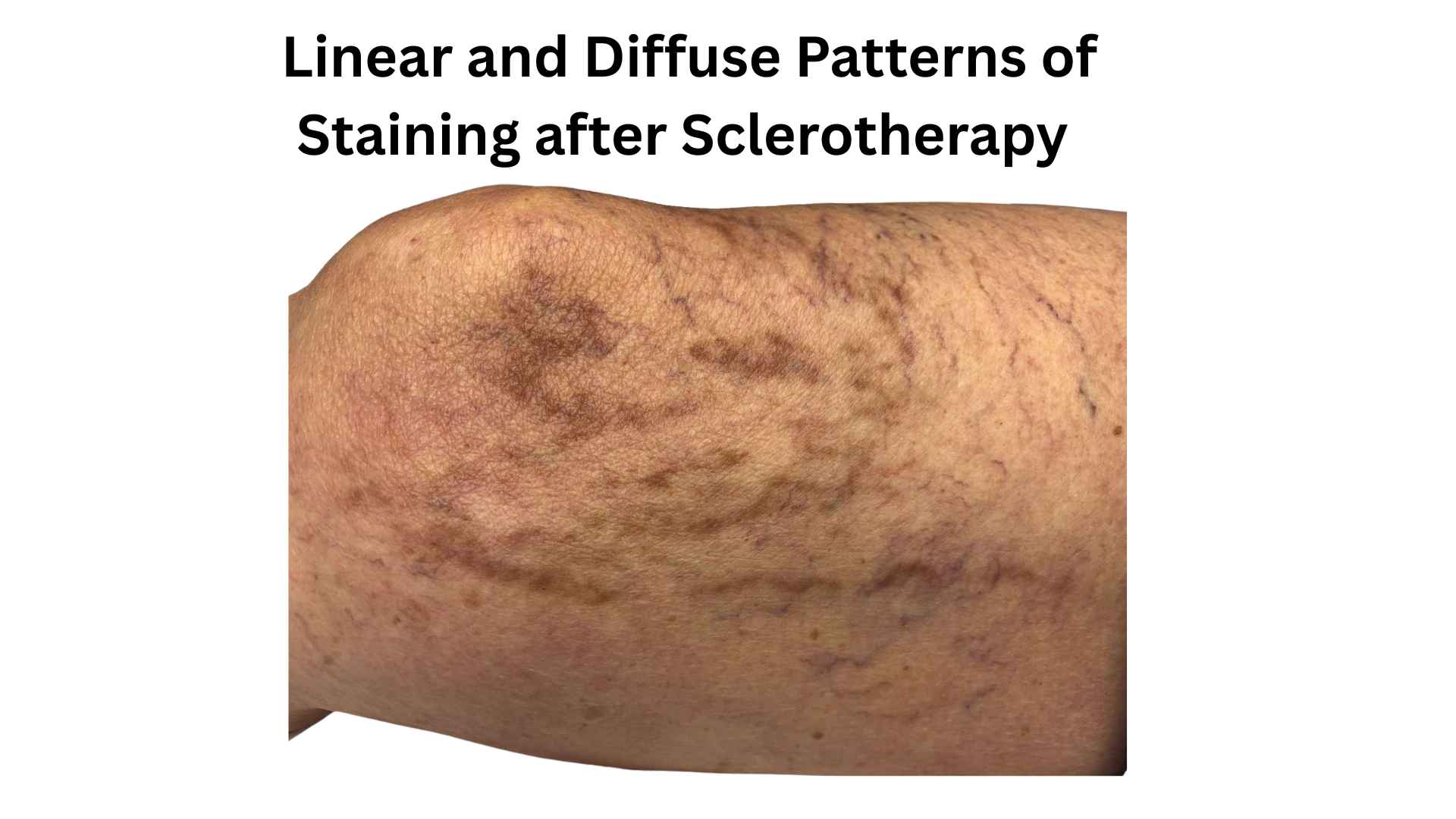
Linear Staining
Linear staining follows the precise anatomical course of the treated vessel — a dark brown line where the spider vein used to be. It is caused by the degradation of intraluminal thrombus: as the coagulum breaks down, haemosiderin is released and deposits along the vessel lumen [1][8]. This is the most common pattern after microsclerotherapy of spider veins and small reticular veins, and it is the more preventable of the two patterns. Early evacuation of intraluminal coagulum at the 1–2 week review significantly reduces its incidence [8].
Diffuse or Nummular Staining
Diffuse staining produces a broader, cloud-like or coin-shaped (nummular) area of discolouration that does not follow the vessel course. It results from vessel wall disruption — typically caused by excessive sclerosant concentration or volume — leading to widespread extravasation of red cells into the surrounding reticular dermis [5][9]. This pattern is more extensive, more cosmetically distressing, and more likely to persist. Its presence almost invariably indicates that the sclerosant was too concentrated or too much was injected at a single site [19][23].
5. Who Is at Risk? — Risk Factors
Patient-Related Risk Factors
Darker Fitzpatrick skin types (III–VI): melanocytes are more reactive to inflammatory stimuli, increasing the post-inflammatory melanin component of PSH [5][20].
Elevated serum ferritin / high body iron stores: Thibault and Wlodarczyk demonstrated a linear relationship between pre-treatment serum ferritin and PSH occurrence in prospective studies [3][4]. Scott and Seiger (1997) noted this relationship was not always consistent, and its clinical utility as a predictor remains debated [6].
Oral contraceptive pill or hormone replacement therapy: oestrogen stimulates melanocyte activity and increases capillary fragility [5].
Iron supplementation: increases circulating iron available for haemosiderin formation [3].
Superficial spider veins: haemosiderin deposits in the reticular dermis are more visible through thin overlying skin in spider vein territory [1][5].
Sensitive or reactive skin: amplified histamine response increases local inflammatory activity [23].
Procedure-Related Risk Factors
High sclerosant concentration: the single most important and modifiable risk factor, with incidence rising from <12% at 0.25% polidocanol to up to 73% at 1.0% [19].
Foam sclerotherapy for small veins: foam acts as a more potent sclerosant at equivalent concentration. Multiple studies confirm higher pigmentation rates with foam versus liquid for C1 disease [9][19].
Perivascular extravasation: incorrect needle placement leading to sclerosant leaking outside the vessel [5][23].
Large intraluminal coagulum: more haemosiderin substrate for degradation. Coagulum evacuation at 1–2 weeks significantly reduces this risk [8].
Inadequate post-treatment compression: allows blood to re-enter the sclerosed lumen, increasing thrombus burden [11][12].
UV sun exposure in the peri-treatment period: stimulates melanocytes in inflamed skin, amplifying the melanin component [5][20].
6. How to Prevent Post-Sclerotherapy Pigmentation — 6 Evidence-Based Strategies
Strategy 1: Use the Correct Sclerosant Concentration
Use the lowest effective concentration of liquid sclerosant. For spider veins and small reticular veins, liquid polidocanol at 0.5% or lower, or liquid sodium tetradecyl sulphate (STD) at 0.2%, represent appropriate starting concentrations [5][19]. Do not use foam for C1 disease. The JEADV systematic review by Bossart et al. (2023) found a near-linear relationship between polidocanol concentration and PSH incidence across all included studies [19]. Kern et al. (2004) confirmed in a randomised comparison that liquid sclerotherapy produced significantly lower pigmentation rates than foam for telangiectatic leg veins [9].
Strategy 2: Treat Feeder Veins Before Spider Vein Clusters
Treat larger reticular feeder veins before addressing the smaller telangiectatic clusters they supply. This reduces intraluminal filling pressure and the tendency for blood to re-enter treated vessels, thereby limiting thrombus volume and haemosiderin load [5][23].
Strategy 3: Evacuate the Coagulum Early
At the 1–2 week post-treatment review, examine all treated areas for regions that are bulging, tender, or beginning to darken. These are sites of retained intraluminal coagulum. A simple nick with a 19–21G needle followed by gentle expression of the retained blood dramatically reduces the haemosiderin burden and, consequently, the risk of linear staining.
Evidence: Scultetus AH, Villavicencio JL, Kao TC, et al. Microthrombectomy reduces postsclerotherapy pigmentation: multicenter randomized trial. J Vasc Surg. 2003;38(5):896–903. [Ref 8] — multicentre RCT, n=96 patients; significantly reduced PSH at follow-up (p<0.05).
Strategy 4: Prescribe Adequate Graduated Compression
Graduated compression after sclerotherapy reduces the residual vein lumen, promotes apposition of vessel walls, limits the volume of blood available for thrombus formation, and reduces local hydrostatic pressure. Rabe et al. (2007) [11] demonstrated in a randomised controlled study that compression significantly reduced PSH after sclerotherapy of telangiectasias and reticular leg veins. Nootheti, Cadag, Magpantay and Goldman (2009) [12] showed that an additional three weeks of Class I compression (20–30 mmHg) beyond the immediate post-treatment period further reduced pigmentation rates.
Recommendation: Prescribe graduated compression stockings (20–30 mmHg) for a minimum of three weeks after every treatment session [11][12].
Strategy 5: Advise Strict Sun Protection
Patients should avoid direct UV exposure to treated areas for a minimum of 2–4 weeks after treatment and apply broad-spectrum sunscreen (SPF 30–50+) when outdoors. UV radiation stimulates melanocyte activity in already-inflamed skin, amplifying the post-inflammatory melanin component of PSH [5][20]. This is an easily communicated, zero-cost intervention that is frequently omitted from aftercare instructions.
Strategy 6: Consider Pharmacological Adjuncts in High-Risk Patients
Two venoactive agents have Level 1 evidence supporting their use as adjuncts to sclerotherapy to reduce PSH incidence:
Sulodexide: Gonzalez Ochoa et al. (2021) [18] conducted a randomised controlled trial (n=609) demonstrating that oral sulodexide taken for 7 days before and 3 months after sclerotherapy reduced PSH incidence from 14.8% to 8.7% (p=0.01) without affecting clinical vein clearance outcomes.
Micronised Purified Flavonoid Fraction (MPFF / Daflon 1000mg): The VEIN ACT PROLONGED-C1 national multicentre observational programme by Bogachev et al. (2018) [16] found that MPFF 1,000mg/day commenced 2 weeks before and continued 6 weeks after sclerotherapy reduced PSH rates from 41.2% to 33.9% (p=0.034). The anti-inflammatory mechanism of MPFF — including reduction of leucocyte adhesion and improvement of microvascular permeability — was further characterised by Dompmartin et al. (2016) [15] and Bogachev, Boldin and Lobanov (2018) [17].
7. How to Treat Established Post-Sclerotherapy Pigmentation
First principle: 70% of cases resolve spontaneously by 6 months [5]. For pigmentation present for less than 6 months — reassure, enforce sun protection, and watch.
Topical Depigmenting Agents — Targeting the Melanin Component
Topical agents are first-line treatment for persistent PSH, particularly where the post-inflammatory melanin component is prominent [5][20][23]. They are less effective for pure haemosiderin deposits.
Triple combination cream (Kligman's formula): hydroquinone 4% + tretinoin 0.05% + topical corticosteroid (e.g., mometasone furoate 0.1%). This suppresses melanin synthesis, accelerates epidermal turnover to remove pigmented keratinocytes, and limits irritant reactions. It is the most widely used and evidence-supported topical regimen for post-inflammatory hyperpigmentation.
Azelaic acid (15–20%): inhibits tyrosinase and has cytotoxic effects on abnormally active melanocytes.
Kojic acid (1–4%): a fungal tyrosinase inhibitor, often used in combination formulations.
Topical retinoids (tretinoin, adapalene): accelerate epidermal cell turnover and disperse melanin granules.
Topical vitamin C (ascorbic acid): antioxidant and tyrosinase inhibitor; reduces oxidative conversion of tyrosine to melanin.
Deferoxamine Mesylate — Targeting the Haemosiderin Component
Deferoxamine mesylate (DFO) is an iron-chelating agent that directly targets the haemosiderin component of PSH — the mechanism that topical agents cannot address. The concept of topical iron chelation for varicose pigmentation was first described by Myers in 1966 [2].
Subcutaneous injection: Lopez, Dilley and Henriquez (2001) [7] demonstrated that weekly subcutaneous DFO 500mg injections achieved 81–100% depigmentation in 27–46 days in treated patients, compared to 150–255 days in untreated controls — an 82% reduction in time to resolution (p<0.001).
Intradermal mesotherapy: Mello Netto, Zeilmann, Santos and Corassa (2025) [21] described monthly intradermal DFO 50mg/mL mesotherapy in a case series achieving complete clearance in the majority of patients within 3–4 sessions. The intradermal route targets haemosiderin at the level of the reticular dermis where it is deposited.
Laser and Light-Based Treatments — Targeting Both Pigments
Energy-based devices are the gold standard for persistent PSH that has not responded to topical or chelation treatments [14][22][23].
Q-switched Nd:YAG laser (1064nm): causes selective photoacoustic fragmentation of both haemosiderin and melanin granules. Macrophages then transport the smaller fragments via lymphatics. Multiple published series report improvement in over 90% of patients.
Intense Pulsed Light (IPL) ± radiofrequency: Mlosek, Wozniak, Malinowska et al. (2012) [14] reported complete regression in 90.5% of 21 patients, with ultrasound-confirmed normalisation of dermal echogenicity.
Combined laser approaches (Nd:YAG + Er:YAG + IPL): documented in a 2025 case report by Alekseev and Mishanina [22] for complex post-sclerotherapy pigmentation following Nicolau syndrome.
8. Management Algorithm
0–6 weeks: Reassurance. Sun protection (SPF 30–50+). Compression. Coagulum evacuation if indicated at 1–2 week review [8]. Topical vitamin C or antioxidant serum.
6 weeks – 6 months: Continue above. Add topical depigmenting agents: hydroquinone, kojic acid, azelaic acid, tretinoin. Strict photoprotection essential [5][23].
6–12 months (persistent): Triple combination cream (hydroquinone 4% + tretinoin 0.05% + corticosteroid). Consider deferoxamine mesylate (subcutaneous or intradermal) [7][21]. Laser/IPL evaluation.
> 12 months (resistant): Q-switched Nd:YAG laser ± IPL [14]. Deferoxamine mesotherapy [21]. Combined approaches targeting both haemosiderin and melanin [22]. Identify dominant pigment before selecting modality [20].
9. Key Takeaways for Practitioners
PSH is common (10–30% short-term [5]) but largely preventable — sclerosant concentration is the most controllable risk factor [19].
Two distinct pigments are responsible: haemosiderin (all cases [1][20]) and post-inflammatory melanin (a significant subset [20]). Both must be considered.
Use liquid polidocanol ≤0.5% for spider veins. Avoid foam for C1 disease [9][19].
Evacuate intraluminal coagulum at the 1–2 week review — this is the single highest-impact preventive intervention [8].
Prescribe 3 weeks of graduated compression (20–30 mmHg) after every session [11][12].
Advise strict UV avoidance for 2–4 weeks post-treatment [5][20].
Consider sulodexide [18] or MPFF [16][17] as adjuncts in higher-risk patients.
For established PSH: topical agents for melanin, deferoxamine for haemosiderin [7][21], laser for both [14][22].
Reassure patients: 70% of cases resolve spontaneously by 6 months [5].
🎥 Watch the full 12-minute video covering all of these topics: "Brown Marks After Microsclerotherapy — What Every Practitioner Needs to Know" → [https://youtu.be/ave538uC8xo]
📖 This article is part of the VeinCare Academy clinical education series for healthcare professionals performing microsclerotherapy. Visit veincare.academy for more.
10. References
1. Goldman MP, Kaplan RP, Duffy DM. Postsclerotherapy hyperpigmentation: a histologic evaluation. J Dermatol Surg Oncol. 1987;13(5):547–50. doi:10.1111/j.1524-4725.1987.tb00940.x. PMID: 3571692
2. Myers HL. Topical chelation therapy for varicose pigmentation. Angiology. 1966;17(1):66–8. doi:10.1177/000331976601700109
3. Thibault P, Wlodarczyk J. Postsclerotherapy hyperpigmentation: the role of serum ferritin levels and the effectiveness of treatment with the copper vapor laser. J Dermatol Surg Oncol. 1992;18(1):47–52. doi:10.1111/j.1524-4725.1992.tb03300.x. PMID: 1740567
4. Thibault PK, Wlodarczyk J. Correlation of serum ferritin levels and postsclerotherapy pigmentation: a prospective study. J Dermatol Surg Oncol. 1994;20(10):684–6. doi:10.1111/j.1524-4725.1994.tb00453.x. PMID: 7930015
5. Goldman MP, Sadick NS, Weiss RA. Cutaneous necrosis, telangiectatic matting, and hyperpigmentation following sclerotherapy: etiology, prevention, and treatment. Dermatol Surg. 1995;21(1):19–29. doi:10.1111/j.1524-4725.1995.tb00107.x. PMID: 7600016
6. Scott C, Seiger E. Postsclerotherapy pigmentation: is serum ferritin level an accurate indicator? Dermatol Surg. 1997;23(4):281–3. PMID: 9149795
7. Lopez L, Dilley RB, Henriquez JA. Cutaneous hyperpigmentation following venous sclerotherapy treated with deferoxamine mesylate. Dermatol Surg. 2001;27(9):795–8. doi:10.1046/j.1524-4725.2001.01052.x. PMID: 11553167
8. Scultetus AH, Villavicencio JL, Kao TC, et al. Microthrombectomy reduces postsclerotherapy pigmentation: multicenter randomized trial. J Vasc Surg. 2003;38(5):896–903. doi:10.1016/s0741-5214(03)00920-0. PMID: 14603191
9. Kern P, Ramelet AA, Wutschert R, et al. Single-blind, randomized study comparing chromated glycerin, polidocanol solution, and polidocanol foam for treatment of telangiectatic leg veins. Dermatol Surg. 2004;30(3):367–72. doi:10.1111/j.1524-4725.2004.30102.x. PMID: 15008862
10. Guex JJ, Allaert FA, Gillet JL, Chleir F. Immediate and midterm complications of sclerotherapy: report of a prospective multicenter registry of 12,173 sclerotherapy sessions. Dermatol Surg. 2005;31(2):123–8. doi:10.1111/j.1524-4725.2005.31027. PMID: 15762201
11. Rabe E, Schliephake D, Otto J, et al. Compression after sclerotherapy for telangiectasias and reticular leg veins: a randomized controlled study. J Vasc Surg. 2007;45(6):1212–6. doi:10.1016/j.jvs.2007.02.039. PMID: 17467226
12. Nootheti PK, Cadag KM, Magpantay A, Goldman MP. Efficacy of graduated compression stockings for an additional 3 weeks after sclerotherapy treatment of reticular and telangiectatic leg veins. Dermatol Surg. 2009;35(1):53–7. doi:10.1111/j.1524-4725.2008.34382.x. PMID: 19067733
13. Guex JJ. Complications of sclerotherapy: an update. Dermatol Surg. 2010;36 Suppl 2:1056–63. doi:10.1111/j.1524-4725.2009.01409.x. PMID: 20891036
14. Mlosek RK, Wozniak W, Malinowska S, et al. The removal of post-sclerotherapy pigmentation following sclerotherapy alone or in combination with crossectomy. Eur J Vasc Endovasc Surg. 2012;43(1):100–5. doi:10.1016/j.ejvs.2011.10.005. PMID: 22078293
15. Dompmartin A, Le Touze A, Grenier N, et al. Beneficial effects of the micronized purified flavonoid fraction (MPFF, Daflon® 500mg) on microvascular damage elicited by sclerotherapy. Phlebology. 2016;31(1):50–6. doi:10.1177/0268355514564414. PMID: 25514922
16. Bogachev VY, Boldin BV, Turkin PY. Administration of micronized purified flavonoid fraction during sclerotherapy of reticular veins and telangiectasias: results of the national, multicenter, observational program VEIN ACT PROLONGED-C1. Adv Ther. 2018;35(7):1001–1008. doi:10.1007/s12325-018-0731-z. PMID: 29949043
17. Bogachev VY, Boldin BV, Lobanov VN. Benefits of micronized purified flavonoid fraction as adjuvant therapy on inflammatory response after sclerotherapy. Int Angiol. 2018;37(1):71–78. doi:10.23736/S0392-9590.17.03868-8. PMID: 28945060
18. Gonzalez Ochoa AJ, Carrillo J, Manríquez D, et al. Reducing hyperpigmentation after sclerotherapy: a randomized clinical trial. J Vasc Surg Venous Lymphat Disord. 2021;9(1):154–162. doi:10.1016/j.jvsv.2020.06.019. PMID: 32739509
19. Bossart S, Daneluzzi C, Cazzaniga S, Ramelet AA, Uthoff H, Seyed Jafari SM, Baumgartner M, Hunger RE, Heidemeyer K, Willenberg T. Skin hyperpigmentation after sclerotherapy with polidocanol: a systematic review. J Eur Acad Dermatol Venereol. 2023;37(2):274–283. doi:10.1111/jdv.18639. PMID: 36196455
20. Bossart S, Ramelet AA, Seyed Jafari SM, Yawalkar N, Feldmeyer L, Heidemeyer K. Revealing the origin of postsclerotherapy hyperpigmentation: identification of melanin and hemosiderin as causative pigments in a histopathological study. Phlebology. 2025 Jun 30:02683555251357095. Online ahead of print. doi:10.1177/02683555251357095. PMID: 40586436
21. Mello Netto BAS, Zeilmann E, Santos GS, Corassa JM. Treatment of hyperpigmentation after sclerotherapy through mesotherapy with deferoxamine mesylate: a case series. Cureus. 2025;17(12):e98954. doi:10.7759/cureus.98954. PMID: 41523378
22. Alekseev N, Mishanina V. Combined laser therapy for post-sclerotherapy hyperpigmentation following Nicolau syndrome: a case report. Cureus. 2025;17(4):e82375. doi:10.7759/cureus.82375. PMID: 40385807
23. Bentivedo A, De Ruggieri G, Floris M, Mastrangeli MR. When sclerotherapy leaves its mark: prevention and management of cutaneous complications. Veins and Lymphatics. 2026;15(1). doi:10.4081/vl.2026.14912
© VeinCare Academy | Dr. Haroun Gajraj | veincare.academy
This article is intended for qualified healthcare professionals. All clinical decisions should be based on individual patient assessment and current professional guidelines.
© VeinCare Academy | Dr. Haroun Gajraj | veincare.academy
This article is intended for qualified healthcare professionals. All clinical decisions should be based on individual patient assessment and current professional guidelines.
About the author
This educational article is written and regularly reviewed by Dr Haroun Gajraj, a GMC‑registered vein specialist who has treated thousands of patients with vein disease and has trained many doctors and nurses in microsclerotherapy, shortwave diathermy and related cosmetic vein procedures.
Dr Gajraj is the founder and board member of the British Association of Sclerotherapists.
You can view his current GMC registration and independent patient reviews on iWantGreatCare for further information about his clinical background. It is designed for healthcare professionals and is based on current clinical guidelines, peer‑reviewed research and day‑to‑day practice experience. The information here is general education only and is not a substitute for individual clinical judgement, local protocols or formal training. Clinicians remain responsible for assessing each patient, obtaining informed consent, explaining risks and alternatives, and working within the scope of their professional registration and regulatory guidance.
Subscribe
For regular updates get my weekly newsletter here