- Jul 15
Why the Ankle is a Bad Place to Start Learning Microsclerotherapy
- Haroun Gajraj
- Microsclerotherapy
By Dr. Haroun Gajraj | VeinCare Academy | 15th July 2026
Written by Dr. Haroun Gajraj, GMC-registered Vascular Specialist, Founder & Board Member of the British Association of Sclerotherapists (BAS).
This review is written for healthcare professionals who are learning or performing Microsclerotherapy for leg telangiectasia. It looks specifically at spider veins around the ankle, why this site behaves differently from the rest of the leg, and why it is a poor place for a beginner to start injecting.
Key Clinical Takeaways (TL;DR)
Ankle spider veins are not usually cosmetic: A fan-shaped pattern here often represents corona phlebectatica, a sign of advanced chronic venous disease rather than simple C1 telangiectasia.[3][4]
Formal CEAP C4c classification: Since 2020, corona phlebectatica is classified as C4c disease, explicitly shifting it out of the cosmetic category.[2]
High correlation with underlying reflux: The presence of an ankle flare correlates with a 4.4-fold higher relative risk of incompetent leg or calf perforators.[3]
Duplex ultrasound is strongly recommended: ESVS 2022 guidelines recommend duplex ultrasound before treating telangiectasias, so that significant proximal reflux is identified and addressed first. This is a Class I recommendation, though based on Level C (expert consensus) evidence rather than randomised trial data.[1]
Narrow technical margin for error: The ankle has a higher density of superficial neurovascular structures than the mid-calf or thigh, which is a teaching point grounded in anatomy rather than a quantified trial-based complication estimate.
The practical default for beginners: Novice practitioners should build technical mastery and diagnostic pattern recognition on the thigh and calf before attempting ankle injections.
Contents
Introduction
Why the Ankle is Not Just Another Patch of C1 Disease
What Corona Phlebectatica Tells Us: The Hemodynamic Evidence
Why Beginners Specifically Should Be Cautious Here
What the Guidelines Say: ESVS 2022 Framework
The Evidence Base, and What is Missing From It
A Practical Clinical Policy for Ankle Presentations
Key Clinical Points
Frequently Asked Questions (FAQs)
References
🎥 Prefer video? Watch the full review on YouTube: "Why You Should NEVER Inject Ankle Spider Veins (As a Beginner)" [ click here ]
1. Introduction
Most spider veins on the lower limb are straightforward CEAP C1 disease: intradermal telangiectasias under 1 mm that respond predictably to standard Microsclerotherapy technique. Around the ankle, that assumption needs to be checked before a needle goes near the skin.
A cluster of fine vessels at the medial or lateral malleolus can be an entirely ordinary cosmetic presentation. It can also be corona phlebectatica, a recognised cutaneous sign of chronic venous disease that carries its own classification, its own associations with underlying reflux, and its own reason for pausing before treatment.[2][3]
For an experienced practitioner, distinguishing between the two is part of routine clinical assessment. For a beginner, the visual overlap makes misdiagnosis easy. This review sets out why the ankle deserves a different starting assumption from the rest of the leg, and why it is a sensible site to leave until later in a practitioner's development.
2. Why the Ankle is Not Just Another Patch of C1 Disease
Spider veins are usually grouped, without much thought, into CEAP C1: uncomplicated telangiectasia and reticular veins. That grouping holds up well on the thigh, the calf, and most of the lower leg.
At the ankle and foot, a different pattern can appear: fan-shaped intradermal telangiectases, often accompanied by venous cups and stasis spots. This is corona phlebectatica, classically described around the medial and sometimes lateral malleolus.[3][4] It is not a cosmetic curiosity. The 2020 update to the CEAP classification formally added corona phlebectatica as the C4c subclass, moving it out of C1, and the ESVS 2022 guidelines adopted this staging.[1][2]
Once a practitioner recognises the corona pattern, the case has moved out of simple cosmetic C1 territory and into a chronic venous disease assessment. This is important clinically because C1 and C4c disease call for different management strategies. While C1 telangiectasias can be treated on their own merits in appropriately selected patients, C4c disease signals that a proximal abnormality (superficial reflux, perforator incompetence, or both) is causing what looks like a simple surface finding.
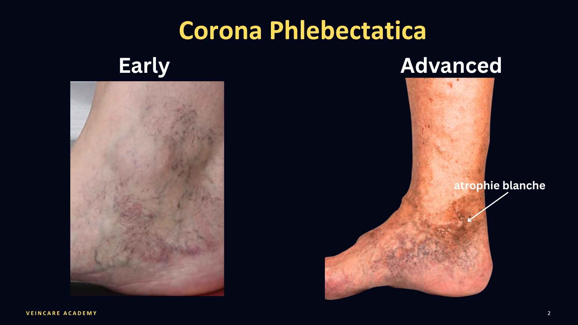
3. What Corona Phlebectatica Tells Us: The Haemodynamic Evidence
The strongest evidence for the clinical significance of corona phlebectatica comes from Uhl and colleagues, who collected full clinical and duplex records on 287 patients across ten European countries.[3] The presence of corona and its severity significantly with advanced CEAP severity, venous symptoms, superficial reflux in both saphenous and non-saphenous territories, and, in particular, perforator incompetence. Patients with corona had a 4.4-fold higher relative risk of incompetent leg or calf perforators on duplex. Deep venous reflux was not an association in that cohort.[3]
A subsequent analysis by the same group in 262 unselected patients (524 limbs) identified which specific components of the corona pattern carried the most diagnostic weight. Blue telangiectases and stasis spots were found to be the most significant components for advanced venous insufficiency.[4] A separate small physiological study using laser-Doppler assessment also found abnormal resting flux and amplitude patterns in corona skin compared with controls, which is consistent with chronic disease, though this is a single small observational study rather than confirmatory evidence.[7]
Taken together, the evidence tells us that an ankle telangiectatic flare is not something to assess by visual inspection and then treat without investigation. It is a surface manifestation of underlying perforator or superficial reflux that should be identified before planning treatment.
4. Why Beginners Specifically Should Be Cautious Here
Two distinct issues make the ankle a higher-risk zone for novice injectors:
1. The Diagnostic Problem
Distinguishing isolated, cosmetic C1 ankle telangiectasia from an early corona phlebectatica is difficult even for very experienced clinicians. The general consensus is that telangiectasia around the ankle should be investigated by duplex ultrasound.
2. The Anatomical Margin of Error
As emphasised in VeinCare Academy training courses, the ankle carries a higher density of superficial neurovascular structures than the mid-calf or thigh, and the skin is thin and tightly bound over bony prominences, which narrows the technical "margin for error". When learning Microsclerotherapy, there is a tendency in the early stages to inject too deeply. This exposes the underlying neurovascular bundle to inadvertent injury or injection. accidental injection of sclerosant into an artery. It is a catastrophe. In our courses, we advise trainees not to inject the ankle area until they have built up considerable experience and not until they have refined their injection technique It is worth being clear that this anatomical caution is a teaching point grounded in anatomy and training experience, not a quantified trial-based complication estimate.
Put these two factors together, and the beginner's instinct to treat a straightforward-looking ankle vein exactly like a C1 vein on the thigh or calf is understandable.
For beginners, the advice is clear: do not inject veins around the ankle
5. What the Guidelines Say: ESVS 2022 Framework
The European Society for Vascular Surgery (ESVS) 2022 Clinical Practice Guidelines give the most direct guidance on available, though it is worth remembering that the specific recommendations below are Level C: based on expert consensus rather than randomised trial data.[1]
Duplex First: For all patients presenting with reticular veins or telangiectasias around the ankle, ESVS recommends duplex ultrasound of the lower limb veins before treatment to look for associated incompetent veins (Class I, Level C).[1]
Reflux First: The guidelines recommend that significant associated incompetent veins be treated first, before smaller surface veins are addressed (Class I, Level C).[1]
Selective Local Treatment: ESVS 2022 upgraded sclerotherapy for reticular veins to first-choice treatment (Class I, Level A), while transcutaneous laser was upgraded as a treatment option for telangiectasias specifically (Class IIa, Level B). Foot and ankle varicose veins and telangiectasias specifically may be considered for local treatment during or after ablation of proximal reflux (Class IIb, Level C).[1]
Together, this guidance supports a duplex-first and reflux-first strategy for the ankle, with local intervention permitted once larger haemodynamic problems have been excluded or addressed. No dedicated NICE, SIGN, or equivalent guideline specifically addressing ankle spider vein injection was identified for this review; ESVS 2022 is the guideline body that speaks most directly to this site.[1][8][9]
6. The Evidence Base, and What is Missing From It
No randomised or prospective comparative study specific to ankle spider vein injection was identified. Decisions about ankle injection therefore rely on disease classification, duplex findings, and guidelines rather than on direct trial evidence for the ankle itself.

The absence of trials related directly to the ankle is an important finding. It means a decision to inject an ankle vein rests on guidelines and disease classification rather than on direct trial evidence that ankle injection alone is safe, durable, or comparable to a staged approach.[1][5]
7. A Practical Clinical Policy for Ankle Presentations
Duplex Investigation IS Indicated When:
The telangiectatic pattern is perimalleolar, fan-shaped, or clustering around the malleoli.[3][4]
There are accompanying symptoms, local oedema, visible reticular varicosities, or early skin discoloration.[1]
Clear CEAP staging is required to differentiate whether the presentation is a benign cosmetic C1 or a pathologic C4c corona pattern.[1][2]
Deliberate assessment of superficial reflux and perforator competence is required to safely sequence care.[3]
Duplex Investigation is NOT Indicated As Routine When:
A comprehensive lower limb duplex scan has already been performed, and findings give no reason to suspect proximal obstruction, deep venous insufficiency, or mixed truncal disease.[1]
Local Microsclerotherapy IS Indicated When:
A duplex ultrasound has excluded significant associated proximal or perforator incompetence, or relevant proximal reflux has already been treated.[1]
Local ankle treatment is being delivered as a staged refinement during or after successful treatment of proximal reflux (ESVS Class IIb, Level C framework).[1]
The patient has been explicitly informed that while general telangiectasia sclerotherapy evidence is strong, ankle-specific efficacy and safety data is limited.[5][6][10]
The procedure is performed by a clinician experienced in both venous assessment and micro-injection technique.
Local Microsclerotherapy is NOT Indicated When:
Duplex ultrasound reveals significant, untreated superficial or perforator reflux upstream.[1]
The pattern is suspicious for corona phlebectatica, and formal venous assessment or CEAP staging has not yet been completed.[1][2][3]
The patient expects guaranteed cosmetic clearance without understanding that underlying venous hypertension may cause early recurrence or hyperpigmentation.[3][5]
8. Key Clinical Points
Ankle spider veins need a second look. They cannot automatically be assumed to be cosmetic C1 disease. A fan-shaped, perimalleolar pattern is often corona phlebectatica (CEAP C4c).[2][3][4]
Correlates with underlying reflux. Corona phlebectatica correlates with superficial and perforator reflux, with a 4.4-fold higher relative risk of incompetent perforators in one large cohort.[3]
Duplex is important. ESVS 2022 supports a duplex-first approach. Significant proximal incompetence is generally addressed before local ankle injections.[1]
No dedicated ankle trial data. There is no direct randomised evidence on whether isolated ankle spider vein injection is safe, effective, or durable without addressing proximal disease.[1][5]
Novices should build experience elsewhere first. Differentiating cosmetic C1 veins from C4c disease takes pattern recognition built over case volume. Combined with the anatomical margin for error at the ankle, this is a site best left until later in training.
9. Frequently Asked Questions (FAQs)
Q: Are all ankle spider veins classified as corona phlebectatica? A: No, not all ankle spider veins are corona phlebectatica. Many are simple, cosmetic CEAP C1 telangiectasias. Any cluster around the ankle warrants a deliberate clinical assessment rather than an automatic assumption either way.[2][3]
Q: Does a patient with ankle spider veins always need a duplex ultrasound? A: ESVS 2022 recommends duplex ultrasound before treating telangiectasias generally, and this carries particular weight at the ankle given the documented association between corona and perforator incompetence.[1][3] This is a Class I recommendation based on Level C (expert consensus) evidence, not a randomised-trial-proven requirement.
Q: Can a beginner perform Microsclerotherapy on the ankle? A: It is sensible for beginners to avoid injecting the ankle during early training. Differentiating cosmetic C1 veins from C4c disease takes clinical pattern recognition, and the local anatomy leaves a narrower technical margin for error.
Q: If the duplex scan is completely normal, is ankle injection then straightforward? A: It is more defensible, but ankle-specific outcome data is still absent, and the technical margin at this site remains narrower than on the thigh or calf. Experience still matters.[1][5]
Q: What distinguishes corona phlebectatica clinically from simple C1 disease? A: A fan-shaped distribution around the medial or lateral malleolus, often with blue telangiectases, stasis spots, and venous cups, distinguishes corona from an isolated cosmetic cluster of thread veins.[3][4]
10. References
de Maeseneer MG, Kakkos SK, Aherne T, et al. Editor's Choice: European Society for Vascular Surgery (ESVS) 2022 Clinical Practice Guidelines on the Management of Chronic Venous Disease of the Lower Limbs. Eur J Vasc Endovasc Surg. 2022;63(2):184-267. PMID: 35027279. https://pubmed.ncbi.nlm.nih.gov/35027279/
Lurie F, Passman M, Meisner M, et al. The 2020 update of the CEAP classification system and reporting standards. J Vasc Surg Venous Lymphat Disord. 2020;8(3):342-352. PMID: 32113854. https://pubmed.ncbi.nlm.nih.gov/32113854/
Uhl JF, Cornu-Thénard A, Carpentier PH, Widmer MT, Partsch H, Antignani PL. Clinical and hemodynamic significance of corona phlebectatica in chronic venous disorders. J Vasc Surg. 2005;42(6):1163-1168. PMID: 16376209. https://pubmed.ncbi.nlm.nih.gov/16376209/
Uhl JF, Cornu-Thenard A, Satger B, Carpentier PH. Clinical analysis of the corona phlebectatica. J Vasc Surg. 2012;55(1):150-153. PMID: 21975063. https://pubmed.ncbi.nlm.nih.gov/21975063/
Nakano LCU, Cacione DG, Baptista-Silva JCC, Flumignan RLG. Treatment for telangiectasias and reticular veins. Cochrane Database Syst Rev. 2021;10(10):CD012723. PMID: 34637138. https://pubmed.ncbi.nlm.nih.gov/34637138/
Rabe E, Schliephake D, Otto J, Breu FX, Pannier F. Sclerotherapy of telangiectases and reticular veins: a double-blind, randomized, comparative clinical trial of polidocanol, sodium tetradecyl sulphate and isotonic saline (EASI study). Phlebology. 2010;25(3):124-131. PMID: 20483861. https://pubmed.ncbi.nlm.nih.gov/20483861/
Bihari I, Egresits J, Nemcsik J, Farkas K. Laser-Doppler examination of corona phlebectatica paraplantaris. Int Angiol. 2011;30(2):130-134. PMID: 21427649. https://pubmed.ncbi.nlm.nih.gov/21427649/
Gloviczki P, Comerota AJ, Dalsing MC, et al. The care of patients with varicose veins and associated chronic venous diseases: clinical practice guidelines of the Society for Vascular Surgery and the American Venous Forum. J Vasc Surg. 2011;53(5 Suppl):2S-48S. PMID: 21536172. https://pubmed.ncbi.nlm.nih.gov/21536172/
Wittens C, Davies AH, Bækgaard N, et al. Editor's Choice: Management of Chronic Venous Disease: Clinical Practice Guidelines of the European Society for Vascular Surgery (ESVS). Eur J Vasc Endovasc Surg. 2015;49(6):678-737. PMID: 25920631. https://pubmed.ncbi.nlm.nih.gov/25920631/
McCoy S, Evans A, Spurrier N. Sclerotherapy for leg telangiectasia, a blinded comparative trial of polidocanol and hypertonic saline. Dermatol Surg. 1999;25(5):381-385. PMID: 10469077. https://pubmed.ncbi.nlm.nih.gov/10469077/
About the Author

This educational article is written and regularly reviewed by Dr Haroun Gajraj, a GMC-registered vein specialist who has treated thousands of patients with vein disease and has trained many doctors and nurses in microsclerotherapy, radiofrequency thermocoagulation, and related cosmetic vein procedures. Dr Gajraj is the founder and board member of the British Association of Sclerotherapists. You can view his current GMC registration and independent patient reviews on iWantGreatCare for further information about his clinical background.
This article is intended for healthcare professionals and is based on current clinical guidelines, peer-reviewed research, and day-to-day practice experience. The information here is general education only and is not a substitute for individual clinical judgement, local protocols, or formal training. Clinicians remain responsible for assessing each patient, obtaining informed consent, explaining risks and alternatives, and working within the scope of their professional registration and regulatory guidance.
Subscribe
For regular updates get my weekly newsletter here
© VeinCare Academy | Dr. Haroun Gajraj | veincare.academy
This article is intended for qualified healthcare professionals. All clinical decisions should be based on individual patient assessment, primary medical literature and current professional guidelines.
*All references in this blog have been checked against publicly available sources (for example, PubMed and official guideline websites), but this is an educational blog post, not a peer-reviewed journal article. Minor discrepancies in author lists, page numbers or indexing details may remain, and readers should always refer to the original publications and current clinical guidelines before making clinical decisions.